How To Prevent Workplace Injuries

 Why are workplace injuries so common?

The nature of work is that we are often required to complete the same task for hours. We can also find ourselves faced with time constraints and deadlines that lead to lazy postures and taking shortcuts, simply to get the job done.

How can they be prevented?

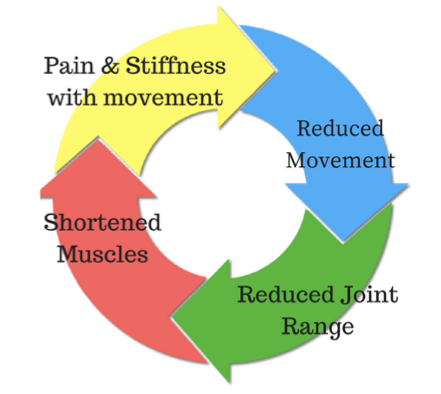
Workplace injuries can happen suddenly, through an accident like a fall or by lifting something too heavy, however, the vast majority of workplace injuries occur over time due to repetitive tasks. Often these conditions begin slowly and take many months to resolve. Here are a few tips to keep yourself pain free in the workplace. 

Moving Items:

It’s important to assess the risk before you start. Do you need to ask for help or use an assistive device? Your legs are the strongest part of your body and ideally, you should use them to power the movement, rather than your arms or back. 

Bending and twisting when lifting is also a common mechanism for injury. It is much safer to lift, then step to turn before putting an object down again. Pushing is a much more efficient movement than pulling and is always preferable if you have a choice. Try to push at waist height and keep forces as close to your body as possible. 

Office Work:

Overuse injuries can occur by using the same side of your body rather than alternating sides. Practise using both left and right hands for taking phone calls and mouse work. 

Be aware of your posture. Good posture isn’t having a rigid and upright spine. It’s about being able to let your spine sit comfortably in its natural curves and be able to move in and out of this easily. Stretching can help to counteract positions you find yourself in for long periods. 

Your physiotherapist is a great person to speak to about preventing injuries in your workplace.

None of the information in this article is a replacement for proper medical advice. Always see a medical professional for advice on your individual injury.

How Vitamin D Can Affect Your Health

 With so much public education about the dangers of sun damage, the last thing you’ll be expecting to hear is that you’re not getting enough sunshine. However, in some countries up to 30% of the population have inadequate vitamin D status, increasing to more than 50% in women during winter and spring. 

What does vitamin D have to do with sunshine? 

Also known as the “sunshine vitamin”, vitamin D is essential for your health. With a typical western diet, most people will get about 10% of their daily vitamin intake through food and the other 90% is processed by the body through exposure to sunshine. 

Why is it important? 

Vitamin D is essential to maintain bone health and muscle function. Deficiencies in children can cause rickets, and over time it can cause osteoporosis in adults. It is also a predictor of falls, due to reduced muscle strength, which coupled with osteoporosis can lead to complicated fractures. Depression has also been linked to vitamin D deficiency. 

Vitamin D has also been shown to have a significant role in preventing respiratory disorders and even reducing their severity. This has been a suggested mechanism for why people with darker skin are more susceptible to COVID infections and suffer more severe infections. 

Think you get enough sunshine? 

You might be surprised at how much sun exposure you need to make enough vitamin D. Unfortunately the answer isn’t straightforward. During winter you’ll need to be in the sun for longer, and the further from the equator you are, the more sun you’ll need. Fair-skinned people are better adapted to process vitamin D and as such need to spend less time outside. 

People who tend to avoid the sun or dress very modestly might be surprised to find that they are vitamin D deficient, along with office workers and those who spend a lot of time indoors, particularly the elderly who are in care. 

Being overweight can also put you at risk of being vitamin D deficient as fat cells absorb vitamin D and prevent it from being released in a way that can be used by the body. Vitamin D deficiency can be diagnosed with a simple blood test. To find out more about how much sun you should be getting and how to balance sun exposure with skin cancer risks go to…. www.sunsmart.com.au/vitamin-d 

None of the information in this article is a replacement for proper medical advice. Always see a medical professional for advice on your individual injury.

MCL Tears

 What is it?

Your knee moves freely backwards and forwards; however, the thought of it moving from side to side probably makes you cringe. This is because the knee joint has sturdy ligaments either side of it that prevent sideways movement and we instinctively know that a lot of force would be required to shift it in this direction. 

The ligaments on either side of the knee are called the Collateral Ligaments and they each work to provide stability and restrict the knee’s movement into a sideways direction. The Medial Collateral Ligament is found on the inside of the knee and act to prevent the knee bending sideways away from the body.

How does this injury occur?

The typical mechanism for this injury is a force that drives the lower leg sideways away from the upper leg. This can occur from an awkward landing from a height, or when twisting with a foot fixed on the ground or from an external force hitting the outside of the knee, such as with a rugby tackle.

What are the symptoms?

MCL tears distinctively create pain and swelling quite specifically on the inside of the knee. The severity of the pain and swelling will be related to the number of ligament fibres damaged. Larger tears may also make the knee feel unstable or loose. 

A grading system is used to classify the severity of the injury and help to guide treatment. Grade 1 indicates that a few ligament fibres have been torn and grade 3 is used for a complete tear of the ligament with associated joint laxity. Very severe MCL tears often also involve injury to the medial meniscus and ACL and can require surgical repair. However, most MCL sprains can be managed well with physiotherapy. Grade 1 and 2 MCL sprains take between 2-8 weeks to fully heal and a complete rehabilitation program is strongly recommended to prevent future injury.  

How can physiotherapy help?

In the early stages of the injury, treatment is focused on pain and swelling management, while allowing the body to start the healing process through inflammation. This is best managed thought the R.I.C.E. principles (Rest, Ice, Compression and Elevation). 

Following any injury, it is natural for muscles to waste a little and the damaged tissues to lose what we call proprioception, the ability to sense their position in space. This loss of muscle strength and proprioception can contribute to further injury if not restored with a proper rehabilitation program. 

Physiotherapy also aims to restore movement to the joint and support the ligament while healing to ensure that it is strong and healthy, and the scar tissue forms in an organized fashion, which makes the new ligament as strong as it can be and protects against future tears.

None of the information in this article is a replacement for proper medical advice. Always see a medical professional for advice on your individual injury. 

Why Do Joints Become Stiff?

 While pain and stiffness often go together, joint stiffness can occur on its own. Joint stiffness can limit your ability to perform usual tasks, for example turning your neck to check behind you while driving. Stiffness can also be a warning sign that part of the body is vulnerable to future injury. There are many different causes of stiffness and we will explore a few of the reasons why you might not be feeling as flexible as normal. 

1. Disuse and lack of movement

Our bodies are made to move. When we are not regularly moving them through their full range, they can begin to feel ‘tight’. This can be caused by a combination of the capsule that surrounds the joint tightening up and the muscles that surround the joint shortening and losing flexibility. Stiff and tight muscles can cause you to feel as though your joints are stiff, even if it is only the muscle length that is restricting the movement. Joint mobilizations, manipulation and muscle stretches/massage can have a significant effect in improving the symptoms.

The most important way to maintain full movement is to regularly move joints through their full range, which also helps to keep muscles and joints healthy. Your physiotherapist can advise you on how to best approach this with a targeted set of exercises.

2. Osteoarthritis (OA)

OA is a degenerative disease, characterized by a breakdown of the joint surface cartilage and the growth of bony osteophytes around areas of stress. While OA is increasingly common as we age, it is thought that the primary cause is abnormal load and stress to joint surfaces and not simply aging itself. As the joint space between two joint surfaces become uneven, joints affected by OA can feel stiff or even ‘blocked’. 

A person with OA will usually feel stiff for around 15-20 minutes after being still. Physiotherapy programs to strengthen the muscles surrounding the joints, so as to help absorb weight-bearing forces, has been shown to have positive results on OA symptoms. 

3. Inflammatory Related Stiffness

The inflammatory process is characterized by swelling and pain around a specific area. Usually, this is a response to damage by the body. As an area swells, this will allow less space for movement and a sensation of stiffness, as anyone who has had sprained an ankle can attest to. Acute inflammation will cause swelling that increases over 24-48 hours and subsides gradually. Autoimmune disorders can cause the body to mistakenly have an inflammatory reaction where there has been no injury, with resulting pain and stiffness. Rheumatoid arthritis and ankylosing spondylitis are two examples of such disorders. 

Stiffness caused by inflammatory disorders is characterized by a feeling of stiffness after rest, particularly in the morning that can take longer than 30 minutes to subside. Inflammatory disorders unrelated to injuries are complex in cause and require collaboration with medical teams for best treatment outcomes. Acute injuries are best managed by following RICE protocols (Rest, Ice, Compression, Elevation).

None of the information in this article is a replacement for proper medical advice. Always see a medical professional for advice on your individual injury. 

Shoulder Labral Tears

 What is the labrum of the shoulder?

The shoulder is a remarkably mobile joint, however, this flexibility comes with the cost of less stability. The glenohumeral joint, where the upper arm meets with the shoulder blade is a ball and socket type joint. The surface area of the ‘socket’ part of the joint (the glenoid fossa) is much smaller than the ball part of the joint (the head of the humerus). A fibro-cartilaginous ring called a labrum surrounds the edge of the glenoid fossa which acts to increase both the depth and width of the fossa. 

This labrum provides increased stability and is also the attachment for a part of the biceps muscle via a long tendon. The labrum can provide flexibility and stability that a larger glenoid fossa might not be able to, however being a soft structure it is prone to tearing which can be problematic. 

What causes the labrum to tear?

The most common way the labrum is torn is through a fall onto an outstretched arm or through repetitive overhead activities such as throwing or painting as the repeated stress on the labrum can cause it to weaken and tear. 

Suspected labral tears can be diagnosed in a clinic by your physiotherapist through a series of tests, however, an MRI is required to fully confirm the presence of a labral tear. Labral tears are classified into different grades, which are determined by their location and severity. This grading is used as a guide to help determine the correct treatment. 

What are the symptoms of a labral tear?

A labral tear is often associated with other injuries, such as rotator cuff tear, which can make the clinical picture a little confusing. Commonly there will be pain in the shoulder that is difficult to pinpoint and the pain will be aggravated by overhead and behind the back activities.

 Severe labral tears can lead to instability and can also be related to dislocations of the shoulder. 

How Can Physiotherapy Help?

The severity and grade of the labral tear will guide treatment. Smaller tears can be treated with physiotherapy that is aimed at increasing strength and control of the shoulder. Other tears may require surgical repair after which physiotherapy is an important part of treatment to rehabilitate the shoulder.  

None of the information in this article is a replacement for proper medical advice. Always see a medical professional for advice on your injury. 

How Do I Know When My Injury Will Heal?

 When injury strikes, the first thing that most of us want to know is ‘how long will this take to heal?’ Unfortunately, the answer to this can be complicated and requires at least a little understanding of how the different tissues of the body heal. Each of the tissues of the body, including muscles, tendons, ligaments and bone, heal at different speeds and each individual will have some variation on those times as a result of their individual health history and circumstances. 

Understanding the type of tissue injured and their different healing times is an important part of how your physiotherapist approaches treatment and setting goals for rehabilitation. On an individual level, a patient’s age, the location and severity of the injury and the way the injury was managed in the first 48 hours all affect the healing times of an injury. Unfortunately, as we age, injuries do tend to heal more slowly than when we are young. Any medical condition that reduces blood flow to an area, such as peripheral vascular disease, can also reduce the body’s ability to heal at its usual rate. 

There are some guidelines that can be followed when predicting how long an injury will take to heal based on the tissue type affected. Muscles are full of small capillaries, giving them a rich blood supply, and as such, they have a comparatively fast healing time with 2-4 weeks for minor tears. This time will be extended for larger tears and more complicated presentations. 

Ligaments and tendons have less access to blood supply and injury to these tissues generally take longer to heal. Larger or complete tears of all soft tissues, may not be able to heal themselves and in rare cases, surgery may be required for complete healing to occur. Similarly, cartilage, the flexible connective tissue that lines the surface of joints is avascular, which means it has little or no blood supply. To heal, nutrients are supplied to the cartilage from the joint fluid that surrounds and lubricates the joint.  

While the different tissues of the body all have different healing times, they do follow a similar process of healing with three main stages, the acute inflammatory phase, the proliferative stage and finally the remodelling stage. 

The inflammatory stage occurs immediately after an injury and is the body’s primary defence against injury. This stage is identifiable by heat, redness, swelling and pain around the injured area. During this phase, the body sends white blood cells to remove damaged tissue and reduce any further damage. This stage usually lasts for 3-5 days. 

The proliferation stage is the phase where the body starts to produce new cells. Swelling and pain subside and scar tissue is formed that eventually becomes new tissue. This stage usually occurs around days 7-14 following an injury. 

The final stage, known as the remodelling stage is when the body completes healing with the reorganisation of scar tissue and the laying down of mature tissue. This stage usually occurs roughly two weeks after the initial injury is sustained. 

At each stage of the healing process, a different treatment approach is required and your physiotherapist can help to guide you through your recovery. Ask your physiotherapist to explain how your injury can be managed best and what to expect in your recovery process. 

Focus on ACL Reconstructions

 A common injury of the knee is a tearing of the ACL (Anterior Cruciate Ligament). This ligament is very important for stability of the knee and often needs to be repaired surgically. The primary function of the ACL is to keep the bottom surface of the knee joint from sliding forwards during movement. An unrepaired knee may feel unstable or give way suddenly.

Not all ACL injuries require surgery and some may heal well with proper rehabilitation, however for those who do need surgery, there is a significant rehabilitation period afterwards. 

What does the surgery consist of?

Every surgeon will have a slightly different technique for surgery. The most common approach is the arthroscopic approach, which uses a small camera and allows the surgeon to make only small incisions into the knee. They will then replace the torn ligament with either a graft from a tendon or ligament at another part of the body or using a synthetic graft.

How long does rehabilitation take?

Full rehabilitation following surgery can take up to nine months and rehabilitation is divided into different stages. As all surgeons will have different protocols for their approaches to surgery, time frames will vary for everyone.  

Initially, after surgery, the graft will be quite weak while a new blood supply is being established. It can take up to 12 weeks before the graft is at its strongest point and evidence shows that it may never have the strength of the original ligament. 

In the early stages, rehabilitation will be focused on restoring movement to the joint and strengthening the muscles around the knee without putting any undue stress on the graft. 

As the graft begins to heal and strengthen, rehabilitation can progress to include stability and control exercises and gradually build up to a complex program that prepares the knee for a full return to sport.

The path to full rehabilitation from a knee reconstruction can be a long and bumpy one, however, there are high success rates with this surgery, particularly when followed up with full physiotherapy rehabilitation.

The information in this article is not a replacement for proper medical advice. Always see a medical professional for assessment of your individual condition. 

Achieving Wellness with Physiotherapy

 While being healthy has always been promoted in society, in recent years, the shift towards wellness has been increasing. While the two concepts seem similar, there are some subtle differences between the two. 

Health refers to the state of wellbeing in physical, mental and social terms, rather than just the absence of disease.  Wellnesshowever, encompasses the ability of a person to experience personal growth in emotional, physical, psychological, spiritual, social and intellectual terms. It can be thought of as thriving rather than just surviving. 

While you most certainly will notice when your health is failing, it can be harder to be aware that you’ve lost a little wellness. When it comes to physical wellness, this may mean that you have no pain, yet still feel a little stiff, or simply can’t do as much as you would like to. 

While in many ways, health can be measured objectively, wellness is a little more subjective. It is determined by your own goals and aspirations. If you are able to participate in activities you love, this can be vital to emotional and social wellness, not only physical health. 

How can physiotherapy help improve wellness?

Your physiotherapist is able to work with you to set goals that are meaningful to you and develop a plan that makes these goals achievable.

Your physiotherapist can also help you to identify factors that may be holding you back from reaching these goals. In some cases, this could be a lack of balance, which reduces confidence in trying something new. Alternatively, an old injury that hasn’t been fully rehabilitated may mean that you are subconsciously avoiding activity.

Little by little, you too can work towards the ultimate goal of total wellness and health. Speak to your physiotherapist about how you can improve your health and wellness.

Focus On Sever’s Disease

 What is Sever’s Disease?

Sever’s disease is a condition that causes pain in the heel of children and adolescents. While it can be quite painful, it is a self-limiting condition that doesn’t usually cause any long-term problems. Sever’s disease is quite common and is the leading cause of heel pain in children and young adolescents. 

Also known as calcaneal apophysitis, Sever’s disease is an inflammation of the growth plate of the heel. A growth plate is the area of bone where new bone is produced in the growing skeleton and is often weaker than other bony areas. Repeated or excessive stress on this area can cause it to become and inflamed and painful.

What are the symptoms?

Children might complain of heel pain that is worse when walking, running or jumping. This may be present following an increase in activity or after a growth spurt. Pain may also be felt when walking on heels.

Why does this happen?

The Achilles tendon attaches to the back of the heel, just next to the growth plate and puts force through this area during gait. If this force becomes too great, this growth plate can become irritated, starting a painful inflammatory process. 

While increased activity is a predisposing factor, other things may also contribute, such as; reduced movement of the ankle, abnormal foot movements when walking and tight calf muscles. It is also possible that Sever’s disease will appear with no apparent cause. 

What is the treatment?

While this is a self-limiting disorder that will go away on its own as the skeletal system completes growth, it can be quite painful and this may impact your child’s activity levels and gait pattern. 

To prevent any long-term issues from adaptations or changes in activity levels, your physiotherapist can work with your child to find solutions that allow maximum movement with minimum pain. Most of the time, simple education and relative rest or ceasing of aggravating activities are effective, however, recent studies have found that properly fitted orthotics can help reduce pain while maintaining activity. Physiotherapists can also address any factors, which may have caused excess stress in this region, such as abnormally tight muscles or poor gait patterns. They can also advise of pain-relieving treatments and alternative forms of exercise if necessary. Speak to your physiotherapist for more information. 

None of the information in this article is a replacement for proper medical advice. Always see a medical professional for advice on your injury.

How Changing Your Posture Can Change Your Life


 Health care professionals seem to constantly be talking about posture. While many people take away the message that they should “stand up straighter” the truth about what good posture is and why you should aim to have it is a little more complicated. 

One of the reasons why posture is so important is that the body has an ideal alignment for almost every joint that provides the most stability and efficiency for movement in that position. 

This is particularly true for the spine, which has a large number of joints that work together to provide movement, stability and support for the body. The spine must also provide a stable base for the shoulder and head. When the spine is in its optimum position, this also allows for free movement of the nerves that supply the trunk, arms and legs.

While the human body is highly adaptable and will continue to function when a posture is not “ideal”, a lot of energy is wasted and undue stress is placed on the muscles, tendons and ligaments of the body. Over time this can cause pain, tightness and loss of flexibility. 

While being able to find these optimum postures is important, it is also important to simply keep moving and not be stuck in the same position for long periods. No matter how ‘ideal’ a posture is, when joints are held in the same position for too long, this can be troublesome. 

Working with a great base posture combined with regular movement and stretches can have a surprising impact on your overall wellbeing. Having good posture has been linked to higher self-esteem, improved concentration, and even better lung function. 

Speak to your physiotherapist for practical tips on how to improve your posture throughout the day.