What Is Frozen Shoulder?

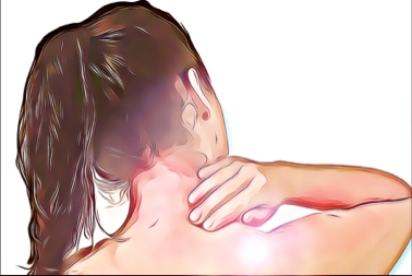
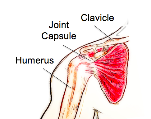
 Frozen shoulder, also known as adhesive capsulitis, is a condition affecting the joint capsule of the shoulder. It is characterized by inflammation of the capsule, leading to pain and stiffness with shoulder movements. 

Frozen shoulder is categorized as either primary or secondary. Primary frozen shoulder occurs for no clear reason, while secondary frozen shoulder develops following an injury or surgery of the joint. 

Frozen shoulder usually follows a typical pattern and can be separated into three stages, freezing, frozen and thawing. The pain begins in the freezing stage as an ache or twinge with movements. The pain gradually increases, and the shoulder begins to feel stiff as well as painful. Usually, shoulder movements away from the body or rotating outwards are the most painful and restricted. 

As the condition progresses, everyday activities can be significantly impacted, with activities such as dressing, grooming, reaching overhead and behind the back becoming difficult. Lifting heavy objects can be very painful, and the pain is often felt at night-time, interrupting sleep. The three stages follow a typical pattern; 

Freezing – Pain is present at rest/night, increasing pain and stiffness with shoulder abduction and external rotation. 

Frozen– Pain starts to lessen, but the stiffness of the shoulder joint increases.

Thawing – Pain reduces to lower levels and movement begins to return.

Frozen shoulder will usually resolve on its own without any long-lasting stiffness. However, complete recovery does not always occur. 

Frozen shoulder usually affects people over the age of 40 and women are affected more often than men. While no definite cause has been identified, there are some factors that increase the risk of developing a frozen shoulder. These include diabetes, prolonged immobilization, trauma, stroke, thyroid dysfunction, heart disease and autoimmune disease.

The early stages of frozen shoulder can mimic other shoulder conditions, and these should first be ruled out by a thorough examination. While frozen shoulder is a self-limiting condition, meaning it will resolve on its own without treatment, this can take up to 2-3 years. Physiotherapy during this time focuses on reducing pain as much as possible and helping patients to cope and adapt to their symptoms during the freezing and frozen stages. 

As the condition moves into the thawing stage, physiotherapy aims to help restore strength, movement and control to the shoulder. The entire process can be extremely distressing for patients and providing support and education as they move through the stages of the condition is an essential part of treatment. 

If you have any concerns about shoulder pain that is not resolving, come and have a chat with one of our physiotherapists to see how we might be able to help you. None of the information in this article is a replacement for proper medical advice. Always see a medical professional for advice on your injury.

Shoulder Instability

 What is it?

Shoulder instability is a term used to describe a weakness in the structures of the shoulder that keep the joint stable, often leading to frequent dislocations. As one of the most flexible joints in the body, the shoulder maintains stability through a balance of support between the dynamic structures (muscles and tendons) and static structures (ligaments and joint shape).

Shoulder instability typically occurs in one of two directions, anterior (forward) or posterior (backwards), anterior instability or dislocations are far more common than posterior. 

What are the symptoms?

The most noticeable symptom of shoulder instability is dislocation or subluxation of the joint. This is often accompanied by pain, clicking sensations, a feeling of instability and in some cases, weakness, tingling, and pins and needles in the arm. Many patients report a feeling of apprehension or instability, as if ‘something is not quite right’. Posterior instability can also cause reduced range of movement and might mimic other common shoulder conditions, which need to be ruled out first. 

How does it happen?

Shoulder instability is also classified as traumatic, occurring after an injury or atraumatic, where the shoulder is exceptionally flexible and prone to dislocations from everyday forces. Instability can also occur from chronic overuse where the shoulder joint is damaged slowly over time.

Traumatic shoulder instability is the most common form. Often the joint is dislocated by a strong force and damaged, leaving it more unstable and vulnerable to future dislocations. Rugby and football players are commonly affected. However, dislocations can occur in the general public from something as simple as falling onto an outstretched hand.

How can physiotherapy help?

Shoulder instability is a complex condition, and each person will have a different combination of causes and structural deficiencies. Physiotherapists are trained to identify issues of coordination, control and strength that may be contributing to instability and provide an extensive rehabilitation program. For some patients, surgery is recommended to help restore some static stability to the joint. However, this is not the best pathway for everyone. If surgery is indicated, a full rehabilitation program is also recommended for the best outcomes. 

Helping patients to understand and manage their condition is an essential part of recovery. Physiotherapy is usually always recommended as the first line of treatment before surgery and can have excellent outcomes, with or without going under the knife. 

None of the information in this article is a replacement for proper medical advice. Always see a medical professional for advice on your injury.

What Is Chronic Ankle Instability?

 Chronic ankle instability, as the name implies, is a chronic condition of instability affecting the ankle and it’s surrounding structures. It usually develops after a severe ankle sprain. However, some people are born with less stable ankles; these individuals are generally extra flexible throughout their bodies. Approximately 20% of ankle sprains lead to chronic ankle instability due to the resulting changes in ligament support, strength, postural control, muscle reaction time and sensation. 

What are the symptoms?

As well as being more susceptible to ankle sprains, people with chronic ankle instability may notice they are extra cautious during high-intensity activities, if running on uneven surfaces or when changing directions quickly. They may experience a sense of weakness or frequent ‘giving way’ when weight-bearing.

What are the causes?

The primary causes of this condition are ligament laxity, decreased muscle strength of the muscles surrounding the ankle and reduced proprioception. 

Following an ankle sprain, ligaments can be stretched and slightly weaker; in severe cases, they have torn altogether, leaving the ankle structurally weaker. Without full rehabilitation, the surrounding muscles also become weaker, and studies have shown that balance and sensation of the ankle can also be reduced. This means that the ankle is more likely to be injured again, creating a vicious cycle leading to further instability.

How can physiotherapy help?

Physiotherapy treatment for chronic ankle instability focuses on improving strength, control and balance with a variety of techniques. This approach can significantly improve ankle stability and reduce the risk of future sprains. Physiotherapists can help patients to regain confidence and get back to their best performance. 

In some cases, orthotic braces for support can be used. However, this can lead to dependence and further loss of strength and control if used unnecessarily. In cases of extreme ligament laxity or if physiotherapy fails, surgery to repair the damaged ligaments is considered. This is usually combined with a full physiotherapy rehabilitation program for greatest success.

If you don’t feel 100% confident with your ankle, come and have a chat with one of our physiotherapist to see if we can help improve your ankle stability. None of the information in this article is a replacement for proper medical advice. Always see a medical professional for advice on your individual injury. 

Plantar Fasciitis

What is it?

Plantar fasciitis is a common condition of the foot and heel affecting both athletes and members of the general public. The plantar fascia is a fibrous band of tissue that attaches to the base of the heel and supports the muscles and arch on the base of the foot. When the plantar fascia becomes chronically irritated, it is referred to as plantar fasciitis. 

What are the symptoms?

Plantar fasciitis is characterised by pain at the base of the heel. The pain is usually noticed upon waking when people take their first steps of the day. The pain usually settles down after walking around, yet may reappear after sitting for a while and getting up again. The pain can usually be reproduced when the inside of the heel is pressed, and the calf muscles might be noticeably less flexible. 

Plantar fasciitis can usually be diagnosed with a physical assessment by a physiotherapist. Left untreated, plantar fasciitis can lead to chronic heel pain, which can have a significant impact on quality of life, interfering with day to day activities. 

What are the causes?

The plantar fascia supports the arches in the foot during weight-bearing and acts as a shock absorber. Small tears can appear in the fascia when it is exposed to excess tension and stress over time. While the exact cause is unknown, there are several risk factors that can increase the risk of this condition developing.

These include obesity, excessive foot pronation, inadequate shoe support, prolonged standing and excessive running. It has previously been thought that plantar fasciitis is linked to or caused by heel spurs. However, this has been shown to be untrue, and many people have heel spurs without any symptoms. 

How can physiotherapy help?

The goal of physiotherapy is to reduce symptoms and support the fascia to reduce and repair any tissue damage. This is done through short term pain reduction strategies such as ice application, rest, activity modification and gentle stretches.

To help reduce the tension on the fascia, lower leg strengthening and balance exercises will be implemented along with orthotics, night splinting and in some cases, corticosteroid injections. A night splint can be helpful in keeping the calf muscles lengthened as they often rest in a shortened position overnight. 

Other treatment options include extracorporeal shockwave therapy and endoscopic plantar release. However, these interventions will also be coupled with physiotherapy treatment for best results. Patients who are not responding to physiotherapy and other conservative management are candidates for surgical release of the plantar fascia. 

None of the information in this article is a replacement for proper medical advice. Always see a medical professional for advice on your individual injury. 

ACL Tears

What is an ACL tear and how does it occur?

The ACL, or anterior cruciate ligament, is a strong piece of connective tissue, which attaches the thigh bone (femur) to your leg bone (tibia). The ACL provides stability to the knee joint as it prevents the tibia from sliding forward relative to the femur.

ACL tears are a common sporting injury; however, can also occur from everyday activities. The most typical mechanisms of injury are landing awkwardly from a jump, twisting the knee, or suddenly stopping while running. The ACL may also be injured during knee hyperextension, or when hit from the outside. Often, other tissues surrounding the knee are also damaged, including the medial collateral ligament, meniscus, joint cartilage, and bone surfaces. The ligament can be stretched, partially damaged or completely torn.

What are the signs and symptoms?

Many people report hearing a “pop” in the knee along with immediate pain and swelling. Decreased range of movement of the knee is common, and the injured knee is typically unable to take full weight when standing or walking. The knee may also feel unstable, with a sensation of “giving way”. Poor balance and coordination may also be experienced. Smaller tears of the ligament may have only mild symptoms, however, more severe tears will have more significant pain, swelling and instability.

Is surgery necessary?

Traditionally, surgery was thought to be necessary for all full-thickness ACL tears. A series of recent studies have shown, however, that outcomes are often the same for people who chose surgery and those who don’t, both in terms of recovery and future risk of osteoarthritis. Individual circumstances will impact this decision; elite athletes and patients with additional meniscal tears often do best with surgery. Generally speaking, however, with time and full rehabilitation, many people can return to their previous levels of activity without surgery. 

How can physiotherapy help?

For both surgical and non-surgical recovery from ACL tears, physiotherapy rehabilitation is essential for a full recovery. Your physiotherapist will assist you with improving your knee’s range of movement, lower limb strength, balance, stability and coordination. You will re-learn the tasks of walking, using stairs, and negotiating obstacles with retraining of your balance and control. 

Early in rehabilitation, the RICE protocol (rest, ice, compress, elevation) is used in conjunction with static resistance type exercises to improve muscle contraction in the leg and increase blood flow in the area.

Throughout your rehabilitation program, you will progress through a variety of strength and mobility exercises targeted towards your individual needs, with goals of returning to your favourite sport or hobby as soon and as safely as possible.

None of the information in this article is a replacement for proper medical advice. Always see a medical professional for advice on your individual injury. 

Move More, Sit Less – 5 Tips From Your Physio

More movement in your day can improve mental, physical health and even workplace productivity. Physiotherapists know that one of the most significant contributors to pain and injury is simply lack of physical activity. Getting more movement in your day doesn’t need to be about high-intensity exercise, all movement can be beneficial. Here are a few tips to help increase the amount of motion in your day. 

1. Try the Pomodoro Technique at work or when studying

The Pomodoro Technique, developed in the late 1980s, recommends breaking work into intervals, usually 25 minutes at a time interrupted by short rests. Studies’ have shown that by creating small time limits, you can focus more intently and the breaks can be useful motivators. 

Use these intervals as prompts for a short walk, some squats or stretches. Not only will you have made your day more productive, but you will also have added some movement to your day. 

2. Challenge a friend to match your steps

A little healthy competition is a great way to keep you motivated to move more. You can set daily, weekly and monthly targets and compare progress to keep you on track. You can use an app like ‘Habitica’ to help track and create movement habits in a game format. 

3. Park further away 

This is an oldie, but a goodie. If you can’t ride or walk to your daily destinations, try parking further away and using the opportunity to walk. Taking the stairs instead of the elevator and getting up and walking while taking phone calls are also great ways to increase your daily movement. 

4. Have a kettle boiling exercise routine.

The time spent waiting for the kettle to boil can feel like an eternity. Use this time to undertake a mini exercise routine. Try fitting in two sets of five squats, five lunges; five heel nurses or try to balance on each leg for two minutes. 

5. Stretch before bed

Gentle stretches before bed are a great way to relax and keep your joints mobile. Create a routine and make it part of our nightly ritual for better sleep and a healthier body. 

Our physiotherapists are happy to help you find strategies to create more movement throughout the day. Come and have a chat with us to see what might work for you.  

Tips For Exercising When You Have Pain

One of the most challenging aspects of living with an injury or chronic pain is how it can quickly impact your exercise routine. If you have been working towards a fitness or weight goal, this can be extremely demoralizing. Here are a few tips that can help to keep you on track while you recover. Staying as active as possible during this time can mean you’re in the best position to reach your goals again once your injury has healed. 

1. Try a new activity. 

When injury strikes, it can be tempting to stop exercising altogether and rest while you recover.  An injury can be frustrating, but it can also be an opportunity to try out a different sport. If you’re a runner with an ankle injury, you can keep up your fitness by swimming instead. Cycling can be an excellent option for people for dealing with knee pain, and if you’re a swimmer with shoulder pain, maybe switch to running for a while. Check with your physiotherapist for some ideas to keep you moving.  

2. Exercise within your limits. 

If you’re getting pain at 5km, this doesn’t always mean you should give up running altogether. Your physiotherapist can help you monitor your symptoms carefully and plan an exercise routine that keeps your fitness up while reducing symptom flare-ups. Staying as active as possible throughout your recovery can also mean that you a better placed to get back to your best performance once symptoms reside.

3. Take the opportunity to improve your footwear and equipment. 

Injury and pain can be a great prompt to look at your equipment and technique. For example, with hip and knee pain, the type of shoes you wear can have a significant difference. Often pain has more than once cause, with technique and equipment more often than not having a substantial impact on the stress placed on your body. Your physiotherapist is an excellent source of advice in this area, don’t hesitate to ask for an assessment. 

4. Take to the water

Hydrotherapy has long been used to help patients with joint pain or muscle weakness exercise. The water helps reduce joint stress and provide extra sensory input that can reduce pain. Exercising in water can be especially helpful for sufferers of chronic pain or those who have pain with weight-bearing.   Speak to your physio for a hydrotherapy program if you’re not sure how to approach exercise in water. 

Our physiotherapist at Cumberland Physiotherapy in Parramatta is happy to discuss your condition with you and share his tips to help you stay pain-free.  None of the information in this article is a replacement for proper medical advice. Always see a medical professional for advice on your condition.

Spinal Stenosis

What is spinal stenosis? 

The spinal cord, nerves and arteries are housed by the spine, which acts as a hard electrical casing to support and protect these vulnerable structures. The spine has a hollow column that allows the spinal cord to run from the brain to the rest of the body. At each spinal segment, nerves exit the spine and supply the tissues of the body. There is also an intricate network of small veins and arteries that provide blood to the spinal cord and vertebrae, providing them with the nutrients needed to operate. 

Spinal stenosis is characterized by a narrowing of the spaces that house the spinal cord, nerves and blood supply. A variety of factors can cause spinal stenosis, however overwhelmingly it is caused by degenerative changes to the spine as we age. Many people over the age of 60 will have spinal stenosis; however, not all will have pain. Clinically, spinal stenosis is used to describe the painful symptoms of this condition rather than just the narrowing itself.

What are the symptoms? 

Pain with walking or standing that radiates into the hips, thighs and even feet is the hallmark of spinal stenosis. Usually, this pain will be reduced with rest and forward movements of the spine. Spinal stenosis is a progressive condition and symptoms will gradually increase over time. The pain is often described as a deep radiating ache and can be associated with fatigue, heaviness, weakness and numbness. It can affect just one leg, however more often will be felt in both legs.  There will often be associated back pain; however, leg pain is usually the most severe complaint. 

How can physiotherapy help?

There are many conditions that need to be excluded before a diagnosis can be made. Your physiotherapist at Cumberland Physiotherapy in Parramatta is able to conduct a thorough examination and accurately diagnose this condition. In some cases, imaging may be requested. As mentioned earlier, many people have stenotic spinal changes without symptoms. Surgery to decompress the restricted nerves and stabilize the spine are used in very severe cases.  

For mild to moderate cases of spinal stenosis, physiotherapy can be extremely beneficial. Your physiotherapist can help you manage your pain through hands-on techniques and by providing a targeted exercise program based on biomechanical assessment. They are also able to help you to understand and manage your day in a way that helps to reduce flare-ups and maintain muscle strength.

If surgery is the right choice for you, your physiotherapist is able to guide you through this treatment pathway, helping you to prepare and recover from surgery to get the best outcome possible. 

None of the information in this article is a replacement for proper medical advice. Always see a medical professional for advice on your condition. 

How To Make The Most Of Your Physiotherapy Treatment

Physiotherapy treatment can be life changing, helping you recover from traumatic injuries, chronic pain and get you on the road to your best performance levels. Here are a few tips to make sure you get the most out of your physiotherapy treatment. 

1. Ask your therapist questions about your injury

Understanding your condition and how to best manage it is one of the most important factors for a successful recovery. Effective therapists allow time for you to ask questions in a non-judgmental environment. There are no stupid questions, if you don’t understand what is happening in your own body it is harder to follow advice and stick to protocols. This can also help you to cope with pain and feel less helpless in your recovery. 

2.  Follow your therapist’s advice and do your exercises

Home exercises are a key part of your recovery, especially when treatment times are limited. Try to stick to your exercise program as seriously as you would a medicine schedule. It is also important to ensure that you are doing your exercises correctly at home. Don’t be afraid to double and triple check your technique before leaving your appointment.

Your therapist will also provide you with advice regarding activities to avoid, how to stretch, when to rest and how to avoid further injury. If you’re not sure about something, ask your therapist to write it down for you. 

3.  Notice your improvements

Nothing can be more disheartening than feeling like the appointments and exercises you’re dilliegently attending to are making no difference. As thereapists, we make regular measurements to track your improvement and know that while  your symptoms might  be staying constant, you are  actually moving more and increasing stress on your body as you recover. Set your own measurements to help you track your recovery. This can help you stick to treatment and feel more positive as you complete your recovery journey. 

4. Set goals and work with your physio to meet these.

The goals of recovery are different for everyone. Some of us want to be able to reach peak performance, such as running a marathon. For others, just getting through the day with a little less pain would be a huge success. Know your own goals and take the time to discuss this with your therapist, who will guide your treatment to help you meet these milestones. 

Matthew is happy to discuss your condition with you and share his tips to help you stay pain-free.  

Wrist Sprains

Wrist sprains are a general term used to describe any injury to the wrist that doesn’t include a fracture. While this can indicate that they are not serious injuries, wrist sprains can be complicated injuries that require supervision and treatment to recover fully. 

The wrist refers to the area where the bones of the forearm, the radius and ulna, meet and join the bones of the hand. The wrist is able to twist on itself and allows the hand to move to face palm up (supination) or palm down (pronation). The hand is also able to move up and down (flexion/extension) and side to side (abduction/adduction). To allow such complicated movements, the joint surfaces of the wrist are held together by a series of ligaments. When a wrist is sprained, it is usually these ligaments that have been damaged. 

What are the symptoms?

The primary symptom of a sprained wrist is pain with movement of the joint or when taking load, such as when holding a heavy object. 

Ligament injuries are given a grading scale to indicate their severity, which can help to guide treatment. Grade I tears refers to a stretching or laxity of the ligament fibers and injuries of this grade usually heal with rest within 2-3 weeks. A grade II classification signifies that there has been a partial tear of the ligament fibers and will often need more time and treatment for recovery. Grade III tears refer to a full thickness rupture of a ligament and may require splinting or even surgery. 

The most common cause of a wrist sprain is a fall onto an outstretched hand. Ligament injuries can also happen gradually through over use, although this is less common. 

What is the treatment?

Your physiotherapist is able to help diagnosis a wrist sprain and can help to rule out a fracture. An X-ray might be required and your physiotherapist will perform special tests to help identify exactly which structure has been injured, giving the injury a grade, to help guide treatment. 

How can physio help?

The key to effective recovery for a wrist sprain is often in ensuring that the right treatment protocols are in place for your injury. Grade I sprains will recover best with gentle exercises and early strengthening while Grade II to III injuries may require splinting or even a surgical consult for repair. 

If surgery is the right course for you, your physiotherapist is able to guide you through this treatment pathway, helping you to prepare and recover from surgery to get the best outcome possible. 

None of the information in this article is a replacement for proper medical advice. Always see a medical professional for advice on your individual injury.