Four Tips For Reducing Knee Pain

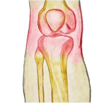
Knee pain comes in many forms with many different causes. While treatment for every person and condition will be different in each case, here are a few tips that may help to reduce knee pain throughout the day.

1. Choose supportive footwear

One of the biggest culprits for ongoing knee pain is wearing unsupportive or high-heeled shoes to work. High heels often lead to tight calves and altered gait patterns, while unsupportive shoes can allow rolling in of the ankles, which can, in turn, place extra stress on the knee joints. Having an assessment with your physiotherapist to see how your shoes might be affecting your knee pain is a worthwhile investment. 

2.  Adjust your sleeping position

While most of the time, our knees get a well-deserved rest during the nighttime hours, there are a few sleeping positions that can place additional stress on the knees. Lying on your side with bent knees can place tension on the outer thigh muscles and also the knee joint itself. Try to keep your knees straightened to at least 30 degrees and if you sleep on your side, place a pillow underneath the top knee to reduce stress on the joint. 

Alternatively, if you sleep on your back it may be helpful to place a pillow under your knees so that they rest in a slightly bent position, to unload the joint.  Try experimenting with different pillow arrangements to see which combination works best for you. 

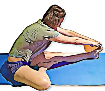
3. Avoid sitting or resting too much

When knee pain strikes, your first instinct is probably to get off your feet and stop exercising. The truth is that our knees, like all our joints, are designed for movement and regular exercise helps to keep them healthy. If you are having pain with high impact activities such as running, try switching to swimming and cycling before stopping exercise altogether. Resting in a sitting position for prolonged periods can also place excess stress over the knee cap and knee joint. When sitting for long periods, try to stretch your legs out ahead of you and avoid crossing your legs.

4.  Seek physiotherapy treatment 

Many of us see putting up with pain as a sign of strength however, a small niggle that is easily treatable can turn into a larger problem if left over time. This may seem like an obvious point, yet the first step to recovery is often just seeking treatment. 

Our physiotherapist is happy to discuss your condition with you and share his tips to help you stay pain-free.

Osteoarthritis of the Knee

Osteoarthritis (OA) is a common degenerative joint disease that affects almost all the joints of the body. The knees are some of the most commonly affected joints, with many people experiencing at least a small degree of osteoarthritis over the age of 40. The disease is characterized by degradation of the cartilage that lines the surfaces of the joint, growth of osteophytes or bony spurs, pain, stiffness and swelling. 

What are the symptoms?

Stiffness in the morning that lasts less than 20 minutes and pain with movement, clicking, crepitus, swelling and a generalized reduction in joint range of motion are all common symptoms of osteoarthritis. As OA is a progressive disease, the condition is categorized into stages to help describe symptoms and guide treatment. Early stages of OA may have only mild symptoms, however as the disease progresses, a joint replacement may be required. 

What causes it?

While aging is the most significant risk factor for the development of OA, it’s not an inevitable outcome of growing older. Other factors that may predict the development of OA are obesity, family history, previous joint injury, high impact sporting activities and peripheral neuropathy. It is thought that abnormal wear and tear or stress on the joint is the primary cause of OA. It is also important to note that many people will have changes on X-Ray that show OA, however, will have no symptoms – which indicates that simply having OA is not a sentence for having pain. 

What is the treatment?

Your physiotherapist is first able to help diagnosis and differentiate OA from other conditions that may have similar symptoms. An X-Ray can confirm the diagnosis and can be helpful in determining the best course of treatment to follow. 

While OA is a progressive disorder, there is often a significant improvement that can be made simply by addressing lifestyle factors and any biomechanical factors that may be contributing to pain. 

How can physio help?

Your physiotherapist is able to guide you with strengthening exercises to support the joint, advice for adapting your exercise routine and can even help you to lose weight, all of which have been shown to have a positive impact on the symptoms of OA. 

If surgery is the right course for you, your physiotherapist is able to guide you through this treatment pathway, helping you to prepare and recover from surgery to get the best outcome possible. 

None of the information in this article is a replacement for proper medical advice. Always see a medical professional for advice on your individual injury. 

Adductor Tendinopathy

What are the symptoms?

The hallmark of this condition is pain in the groin region with movements of the adductor muscles. There may be a feeling of stiffness, weakness and pain when pressing over the adductor tendon. The pain usually begins gradually and progresses over time. It may build up over a few months and may not go away on its own. In severe cases, the pain may impact day-to-day activities, with pain being present when walking or going up and down stairs. Tendon tears may occur suddenly, however tendinopathy is often already present when this happens.

What causes it?

Adductor tendinopathy usually occurs due to chronic overuse, particularly for runners and athletes whose sports involve regular changing of directions. Overstretching of the tendon or an increase in training intensity or type often precede the development of adductor tendinopathies. It is thought that excess forces over an extended period of time cause the tendon tissues to degenerate, becoming painful and more prone to tearing.

What is the treatment?

As many different conditions mimic adductor tendinopathy, accurate diagnosis by a health professional is essential. Certain conditions such as stress fractures of the hip, nerve entrapment or pathologies of the hip should first be ruled out.  

Adductor tendinopathy is treated by first identifying factors that may have led to the development of the condition. Your physiotherapist may recommend a period of rest and suggest that you stop stretching. Common contributing factors are running technique, muscle tightness and/or weakness and training frequency. 

Your physiotherapist is able to help you maintain your training program to the highest level without aggravating your symptoms and help support tendon healing. They are also able to provide support to unload the tendon along with manual therapy and an exercise program, particularly with eccentric exercises, which have been shown to stimulate tendon regeneration.

In most cases, conservative or non-surgical treatment is attempted as the first line of treatment. If this is unsuccessful, cortisone injections are often used to reduce symptoms. In severe cases where the pain persists despite all other attempts at treatment, other medical interventions can be attempted. Once the pain has subsided your physiotherapist is also able to help prevent any further recurrence. 

None of the information in this article is a replacement for proper medical advice. Always see a medical professional for advice on your individual injury.

When Physiotherapy Treatment Doesn’t Work

1. You couldn’t commit to your exercise program.

With our busy modern lives, finding time to make an appointment with your physiotherapist can be hard enough, let alone making time to complete the exercises they prescribe. The tasks set for you at a home by your therapist are often actually more important than the treatment time and are highly targeted for your individual circumstances. Ensuring you are performing your exercises correctly as well as frequently enough are other factors that might leave you seeing little to no improvement. 

2. You weren’t able to continue physiotherapy for long enough. 

While very occasionally, an issue can be resolved in 1-2 visits, most conditions will require at least 5-6 visits for a significant change to be made. Chronic pain and injuries often need much longer still to make an impact. There are many reasons for not being able to return for treatment, however this is one of the most common reasons physiotherapy fails, there’s simply not enough of it to be successful. It is important to discuss with your physio at the beginning of treatment how much time may be needed for a full and effective treatment program. Once

pain and symptoms have resolved, it is also important to complete a full rehabilitation to help prevent future injuries. 

3. Your injury required medical or surgical intervention. 

There are a small percentage of injuries that will require more intensive intervention to heal fully. It is often recomended that physiotherapy be trialled before attempting more invasive treatments. Your physiotherapist and medical team often work together to evaluate your injury and decide the best course of treatment. 

4. Your physiotherapist was simply not the right fit. 

While all physiotherapists are trained to an excellent standard, occasionally you might find that the treatment style of one therapist works better for you than another. It can be worth working with a new therapist before giving up on the idea of physiotherapy altogether. Often physiotherapists within the same clinic are happy to collaborate and offer new perspectives. 

Our physiotherapist is happy to discuss any concerns you have regarding your treatment including issues with previous treatments. None of the information in this article is a replacement for proper medical advice. Always see a medical professional for advice on your individual injury.

Olecranon Bursitis

What is it? 

Bursae are small sacs of fluid found throughout the body. These bursae produce synovial fluid and act to reduce friction between muscles, tendons, ligaments and bones as they move over each other. Bursae are located at strategic points, typically where there are higher points of stress. If a bursa is injured or irritated, it can become inflamed, painful, red and swollen and this condition is referred to as bursitis. 

One bursa that is commonly affected is the olecranon bursa, which sits just over the hard bony process at the base of the elbow. Olecranon bursitis refers to inflammation of the bursa at this point and is a common condition, particularly in men between the ages of 30-60.

What causes it?

Olecranon bursitis has a few different causes including trauma, overuse and infection. A sharp blow to the elbow, through a fall or hit, might damage the bursa leading to bursitis. In other cases, the bursa can be infected by bacteria, which enter the body through a small skin tear. 

Bursitis can also develop slowly through friction of the nearby muscles that cause the bursa to become irritated and inflamed.

What are the symptoms?

The hallmark of this condition is a painful, red, swollen elbow. Typically pain is worst when resting on the tip of the elbow and/or with elbow movements, particularly when bending or straightening the elbow fully. The pain often lasts a few months and may not go away on its own. The pain may build up gradually, or come on suddenly, depending on the cause. Bursitis caused by infection (septic bursitis) may also be associated with general feelings of illness such as fatigue, fever and body aches.

What is the treatment?

As there are many different causes of this condition, accurate diagnosis is essential. Your physiotherapist is able to distinguish between olecranon bursitis and similar conditions such as rheumatoid arthritis or fibromyalgia. Septic bursitis will need to be treated by a medical professional who will determine the best course of action. 

All types of bursitis can be managed initially with a RICE protocol to reduce pain and swelling (Rest, ice, compression and elevation).  Mechanical causes of bursitis can require more in-depth identification of the factors that may have led to the development of this condition. 

Common contributing factors are throwing technique, muscle tightness and/or weakness and training frequency. Your physiotherapist is able to address these factors plus provide taping support to unload the bursa along with manual therapy and an exercise program.

In most cases, conservative or non-surgical treatment is attempted as the first line of treatment. If this is unsuccessful, cortisone injections are often used to reduce pain and inflammation. In severe cases where the pain persists despite all other attempts at treatment, the bursa can be surgically removed in a procedure called a bursectomy. Once the pain has subsided your physiotherapist is also able to help prevent any further recurrence. 

None of the information in this article is a replacement for proper medical advice. Always see a medical professional for advice on your individual injury.

Can Poor Balance Lead To Ankle Sprains?

Ankle sprains are one of the most common sporting injuries and most people have experienced one at least once in their lifetime. While they are common, this doesn’t lessen their negative impacts. Surprisingly, having poor balance might be increasing your risk of ankle sprains. Here we discuss a few facts about balance and what you can do to reduce your risk of ankle injuries.

Why are ankles particularly vulnerable to injuries related to poor balance?

Our ankles have to support our entire body weight when standing on one foot. To provide us with agility as well as stability, our ankles have the ability to move from side to side as well as back and forwards. There is a complicated process constantly operating to keep your foot in the correct position while supporting all this weight, particularly with quick changes of direction, activities done on tiptoes, jumping and landing. 

If the ankle rolls excessively inwards or outwards, the ligaments on the outside of the ankle can be damaged and torn. Balance is an important part of keeping the ankle in the correct alignment and not twisting too far to either side during challenging activities.

A study of high school basketball players by Timothy McGuine et al. in 2010 showed that students with poor balance were up to seven times more likely to sprain their ankle than students with good balance. Other studies have shown that balance training is an effective way of preventing falls in elderly populations. 

Balance can vary from one leg to the other. 

Most of us tend to favour one side of our body for all activities. This is more obvious in the upper body, with most of us identifying as either left or right handed. The same is also true for our lower body, with each of us favouring one leg over the other for balance activities. This can mean that one leg has better balance and strength than the other, leaving the other leg more vulnerable to injury. 

Reduced balance can mean your body has to work harder to perform activities, with muscles activating in a less coordinated way. Improving your balance can also improve your body’s efficiency of movement, which can, in turn, improve your overalls performance without actually improving your muscle strength. 

Balance can be trained rapidly. 

Balance is one of the most overlooked dimensions of physical health however, the good news is that it can be improved relatively quickly. Do a quick check to see if you can stand on each leg for two minutes with your eyes closed. If this is difficult you might find that improving your balance is a great next step in your training program.

Your physiotherapist is able to identify any deficits in your balance is and is able to develop a training program for you to improve your balance. Come and see us for an appointment to see how we can help. None of the information in this article is a replacement for proper medical advice. Always see a medical professional for advice on your individual injury.

Fibromyalgia

What is it? 

Fibromyalgia is a chronic condition characterised by widespread pain throughout the body along with fatigue, memory problems, sleep and mood disorders. Sufferers of fibromyalgia often spend years trying to find a diagnosis that fits their many symptoms and fluctuate between periods of having high energy and ‘crashes’ of fatigue and pain. In severe cases, fibromyalgia can cause significant lifestyle disruptions, including reduced activity, unemployment and depression. 

The underlying mechanism that creates the symptoms of fibromyalgia has been shown to be increased pain amplification by the central nervous system and reduced activation of the sympathetic nervous system. Essentially this means that small pain signals in the body are processed as large pain signals by the central nervous system. 

What causes it?

Fibromyalgia is a complicated condition that is poorly understood. This can be very frustrating for sufferers, who often find themselves being shuffled between health practitioners looking for answers and long term relief. While the pain generally feels muscular, usually little to no muscular damage or injury can be found on physical assessment. The symptoms can also mimic those of an infectious illness, or other chronic diseases. Often a diagnosis of fibromyalgia is reached after other diseases and causes have been ruled out.

The cause of fibromyalgia is as yet unknown, it was thought that the depression and reduced activity that are often associated with fibromyalgia could be causative, however it has been show that these are symptoms of fibromyalgia rather than causes. Other significant signs of fibromyalgia are a lack of REM sleep in sufferers and a positive result of more than 11 out of 18 muscular trigger points. 

What is the treatment?

Following a diagnosis of fibromyalgia, patients primary strategy is to understand and manage their symptoms. This can involve pacing activities and balancing exercise so as to reduce ‘crashes’ and unhelpful pain cycles that lead to frustration. Identifying activities, employment and a routine that don’t exacerbate symptoms can have a significant impact on quality of life for someone with fibromyalgia. Having psychological support can also be very important to help patients deal with the emotional distress of a complex chronic condition that has no outward physical signs. 

Treatments that have been shown to help reduce symptoms are TENS (electrical stimulation) which produces an endorphin response and can reduce pain. Certain medications may be helpful when prescribed by a doctor. Education and understanding of this condition can have the largest impact for sufferers, helping them to manage and maintain some control over their symptoms. Physiotherapists can have a large role in education and helping patients find a routine and activity level that helps them manage their condition as best as possible.

None of the information in this article is a replacement for proper medical advice. Always see a medical professional for advice on your individual injury.

Four Surprising Causes Of Neck Pain

If you are experiencing regular neck pain that just won’t go away, it’s possible that parts of your daily routine are contributing without you realising. Here are a few common everyday activities that might be making your neck pain worse.

1. Your sleeping position

It’s easy to underestimate the impact your sleeping position has however, spending hours in one position will undoubtedly have an effect on your body. Pillows that are too high or too flat can mean your cervical joints are sitting at the end of their range in too much flexion or extension. Similarly, sleeping on your stomach often means your thoracic spine is locked into extension and your neck is fully rotated. In simpler terms, this means your joints are under more stress than necessary. Ideal sleeping posture allows your spine to maintain it’s natural curves.

2. Your daily commute 

Many of us make sure our work stations are ergonomically set up to reduce stress and strain throughout the day. Few of us take the same consideration when it comes to driving. In fact, the set up of your car can be just as important as your work-desk, particularly if you are driving more than 30 minutes everyday. The correct setup in your car can mean you use less effort to drive and turn your head less often to check traffic. 

Ensuring that your steering wheel, seat and mirrors are set up correctly could make a difference to your posture and even perhaps reduce neck pain and headaches. If you find that driving is still affecting your pain after making these changes, try catching public transport or riding a bike on alternative days. 

3. Your downtime 

Many of us unwind by watching TV or our laptops at the end of the day. Your position during this time can be something you give little thought to however, looking up to view a screen mounted on a wall or looking down at a small screen or laptop can put pressure on the upper structures of the neck. Take a few minutes to consider what posture you’re sitting in before settling down to binge watch a series and see if you can either lower the height of your screen or raise it slightly so your neck can be in a more neutral position. 

4. Your exercise routine

Any activity that requires sustained positions or repetitive neck movements can contribute to neck pain. Cyclists can be stuck in neck extension while looking ahead and breast stroke swimmers can also have excess neck extension. Freestyle swimmers with reduced thoracic or neck rotation can have difficulty achieving rotation when breathing which can cause pain and discomfort over time.

Your physiotherapist is able to identify any daily habits or activities that might be contributing to your neck pain. Come and see us for an appointment to see how we can help. None of the information in this article is a replacement for proper medical advice. Always see a medical professional for advice on your individual injury.

Shoulder Labral Tears (SLAP Tears)

What is it? 

A ring of flexible, fibrous connective tissue, known as the glenohumeral labrum, surrounds the shoulder joint. This labrum increases the stability of the shoulder while allowing for the required flexibility of one of the bodies’ most sophisticated joints. One well-known muscle of the arm, the biceps, has an attachment directly into the labrum and is a common site of injury. A tear of the labrum can occur in many locations, however the most common is at the point where the biceps tendon attaches to the labrum. Usually, this tear follows a typical pattern and is referred to as a superior labrum tear, anterior to posterior (SLAP tear). 

What causes it?

SLAP tears can be caused by trauma such as a fall onto an outstretched hand or a dislocated shoulder. Tears can also develop over time with repeated throwing actions or overhead activities as the labrum is weakened and eventually injured. Traumatic tears are more likely to be symptomatic than tears that develop slowly. 

What are the symptoms?

As mentioned, SLAP tears can occur suddenly, through trauma or develop slowly through repeated stress. Often if the injury develops over time, patients can be unaware they have sustained a tear and the injury doesn’t have a significant impact on their pain or function. Preexisting SLAP tears can however, place more tension on the long head of biceps tendon, leading to overuse disorders as a secondary complication.

When the tear occurs through a sudden action or trauma, symptoms can be more marked. Sufferers often notice pain deep in the shoulder joint with overhead shoulder movements, a feeling of weakness, loss of power and/or accuracy with throwing activities. Some people may notice a popping or clicking sensation and occasionally the shoulder may give way. In severe tears, the shoulder might feel unstable and even be at increased risk of dislocation.

How can physiotherapy help?

Your physiotherapist is able to help diagnose a suspected SLAP tear and send you for further imaging if needed. SLAP tears are often graded by severity from I to IV as a way to guide treatment. Physiotherapy is usually recommended as a trial for all tears before considering surgical repair and in many cases can effectively help patients return to their previous activities, symptom-free. 

If physiotherapy is unsuccessful, surgical repair with a full rehabilitation program is recommended. Surgery will usually either repair the tear or reattach the biceps tendon to the humerus (tenodesis). Following surgery, a period of rest in a sling is required before rehabilitation can begin. None of the information in this blog is a replacement for proper medical advice. Always see a medical professional for advice on your individual injury.

Some Surprising Facts About Posture


Your posture is one of the first things other people notice about you and can affect so much more in your life than just spinal health. Healthy posture has been linked to better respiratory health and has even been shown to affect your confidence. In this article, we explore a few other surprising facts about posture. 

Ideal posture doesn’t mean having a straight back.

Many people think having good posture means standing as straight as possible with your shoulders pulled back. This is actually a relatively unnatural posture and requires a lot of energy to maintain. Ideal posture, where the spine rests in it’s most comfortable and strongest position, is where the neck has a gentle curve backwards, the thoracic is curved gently forwards and the lower back curves back again. These curves create an elongated ‘S’ shape and can be seen when viewing a person from side on. 

There are four common postural patterns other than ‘ideal posture’.

For those who don’t have ideal posture, four common variations are seen, these are; kyphosis (excessive curve of the thoracic region), lordosis (excessive curve of the lower back), flat back (loss of the normal spinal curves) and sway back (where the pelvis is pushed in front of your centre of gravity and the upper body leans back to compensate). 

Each of these postures is often accompanied by a typical pattern of joint and muscle stiffness that can lead to pain and injury. Your physiotherapist is able to assess your posture and identify any ways this may be contributing to your pain. They can also help you to change your posture with strategies to increase spinal mobility and strength. 

Regular movement is just as important as your posture.

While ideal posture has been shown to reduce the amount of stress and tension found in the spinal muscles, holding yourself rigidly in one position is also not healthy. Ideally, our spines will be flexible and able to move through their full range without pain or stiffness. Regular movement is the key to healthy joints, including the spine. If you finding yourself sitting or standing for long periods, try to find time to stretch as well as working on your posture. 

Personality type has been linked to postural patterns. A study by S. Guimond and others in 2014 showed a surprising link between personality type and posture. They found that extraverted personalities were much more likely have an ‘ideal posture’ or excessive spinal curves than Introverted personalities and introverted personalities were more likely to have ‘flat’ or ‘sway back’ postures. They were unable to determine if personality influenced posture, vice versa or there was a hidden third factor such as pain.