Three Helpful Stretches For The Busy Office Worker

If you happen to have an office job, it can’t have escaped your notice that sitting has been shown to be associated with a variety of chronic health conditions. Office jobs are one the most common forms of employment in the developed world, so here are some tips to help you stay healthy when sitting all day.


Move more often:

Sitting itself and the posture you find yourself in isn’t as bad as simply being still for hours on end. An expression in physiotherapy is ‘the best posture is your next posture’. This means that, above all, movement is the best thing for your body and those in office jobs can find themselves becoming very still while focused on the next deadline.

Set a quiet alarm to remind you to move or change positions every 20 minutes. Getting up for phone calls and walking over to see colleagues when you have a question is a great way to break up your sitting time.

Reverse your posture:


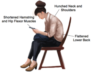
While not moving is definitely the worst aspect to prolonged sitting, the postures we often adopt while sitting can also be problematic. Sitting with a flattened lower back, hunched neck and slouched shoulders is the posture that requires the least energy to maintain and is often the one we sink into in a long day. A slouched posture can lead to shortened hamstring, hip flexor and pectoral muscles.

If you are spending large amounts of time sitting, it’s important to take time every day to adopt the opposite postures and keep your body flexible.
This means moving into thoracic and lumbar extension, stretching your shoulders and extending your hips.

Here are a few stretches you can do every day while seated that will help to reverse your posture. Try to do these stretches every few hours during a working day.


1.Chest stretch


Sit forward, clasp your hands behind your back and lift your arms towards the ceiling. You should feel a stretch at the front of your chest. Look up slightly to increase the stretch. You should not feel any pain or tingling in your arms. Hold for 30 seconds and repeat.

  1. Seated hamstring stretch


Perch on the edge of your seat and straighten one leg out in front of you. Lean forwards at your hips, keeping your back straight. You should feel a gentle stretch at the back of your thigh. If you feel the stretch behind your knee or into your calf, let your ankle relax, and let your foot drop towards the floor. Hold the stretch for 20 second then swap legs, repeat this stretch with each leg twice.

  1. Chin Tuck

Sit up in your chair so your bottom is at the back of your seat and your lower back is supported. Relax your shoulders and gently tuck your chin in, imagine you are holding a soft ball under your chin and are slowly squashing it. You should feel a gentle stretch at the top of your neck. Hold for 20 seconds, release and repeat.

You should not feel any pain with these stretches. Speak to your physiotherapist for a customised stretching routine that you can implement into your day at the office or for more tips on how to perform these stretches to maximum effect. None of the information in this article is a replacement for proper medical advice. Always see a medical professional for advice on your individual condition.

Focus On Anterior Ankle Impingement

What is it?


Anterior ankle impingement, also known as anterior impingement syndrome, is a musculoskeletal condition where repetitive forces compress and damage the tissues at the front of the ankle, causing pain and stiffness. It is a common injury that can affect people of all ages, however is usually seen in athletes of sports involving repetitive or forceful upward movements of the ankle, such as sprinting, landing from long jump, uphill and downhill running.

What are the symptoms?


Pain at the front of the ankle is the primary symptom of anterior ankle impingement. This can be felt as an intense, sharp pain occurring with ankle movements or a dull ache in front of the ankle following periods of exercise. Pain can also be felt when putting weight through the ankle while standing, walking or running. Night-time aching, stiffness, swelling and reduced ankle flexibility are also common symptoms of anterior ankle impingement.

How does it happen?


Anterior ankle impingement is caused by traumatic or repetitive compression to the structures at the front of the ankle as the tibia and talus move towards each other during ankle movements. The tissues that are affected become damaged and inflamed, causing the pain typical of ankle impingement. Chronic inflammation can lead to further stiffness, exacerbating the impingement process.

The most common risk factor for ankle impingement is a previous ankle sprain that was not adequately rehabilitated, as this can result in a stiff or unstable ankle. Another cause of impingement is the growth of small osteophytes or bony spurs around the ankle joint that press against the nearby soft tissues. These can be due to osteoarthritis or grow as a reaction to impingement itself. Training errors, muscle tightness, unsupportive footwear and a hypermobile ankle have also been shown to be risk factors for anterior ankle impingement.


How can physiotherapy help?


Depending on the cause, mild cases of anterior ankle impingement usually recover in one to two weeks with rest and physiotherapy intervention. For more severe impingement, the ankle may require up to six weeks of rest and rehabilitation to recover. In rare cases, surgical intervention will be required to remove any physical causes of impingement, such as osteophytes to restore impingement free movement of the ankle. Your physiotherapist will first identify the cause of your ankle impingement and help you to choose the best course of action to reduce your symptoms. They are able to advise you on the appropriate amount of rest and provide stretches and exercises to restore strength and flexibility to the ankle.

Mobilization techniques and range of motion exercises can also reduce stiffness of the ankle, restoring normal joint movement. Moreover, balance and proprioception exercises are included to prevent further ankle injury. Balance exercises challenge the way your body reacts to outside forces. With this, your balance will be improved, and you’ll have a more stable ankle.

Ideally, physiotherapy treatment is the first step before considering surgery. If surgery is required, your physiotherapist can help you to make a full recovery with a post surgical rehabilitation program.

None of the information in this blog is a replacement for proper medical advice. Always see a medical professional for advice on your individual condition.

Physiotherapy Tips For A More Comfortable Sleep

For most of us, the hours we spend sleeping are simply a time for rest and recovery. However, you might be surprised to learn that your sleeping position can have a significant impact on your body, particularly if you already have an injury. When you consider that we spend approximately 40% of our lives in bed, it becomes less surprising.

Ideally, your body should be held in a position of minimal stress while sleeping. This means that all your joints and muscles are resting in a neutral position. Over time, joints that are held in more extreme positions may put pressure on the surrounding structures and this may lead to a feeling of stiffness in the morning.

Back Pain

For sufferers of back pain, finding a comfortable position at night can be difficult. Ideally, the natural curves of the spine should be maintained and supported throughout the night. The correct mattress will support your lower back without making you feel as though you have been sleeping on concrete all night. A mattress that is too soft might feel comfortable to begin with, but over time will let you sink too much, meaning the curve of the lower spine will be lost. Waking up with a stiff spine could be a sign that you are using the wrong mattress.

For many people, sleeping on their side keeps their spine in a more natural alignment than on their back. If you sleep on your back, placing a pillow under your knees can help to maintain your lumbar spinal curve throughout the night.

Neck Pain


While you may be attached to your pillow, it could be the cause of unnecessary neck pain for you. The neck is often the most vulnerable part of our body when our sleeping setup is not ideal. Side sleepers may let their neck fall excessively to the side with a pillow that is too low or have their neck elevated too much by having their pillows too high.

The importance of having a supportive pillow that supports your neck while sleeping cannot be overstated. If you find yourself putting your arm under your pillow while you sleep, it is likely that your pillow is too low. Having your shoulder in this position overnight can put unnecessary stress on the structures in the shoulder joint and should be avoided if possible.

Sleeping on your stomach with your head turned to the side can be the cause of many issues and if this is your preferred sleeping position, it could be worth chatting to your physiotherapist about strategies improve your sleeping posture.


Hip Pain


Side sleepers often spend their nights with one leg crossed over their body. This can place extra pressure on the structures on the side of the hip, such as tendons and bursa and can impact the health of these tissues as compression can reduce the blood flow to the area. If the mattress is too firm then the hip on the underside of the body may also be compressed under your bodyweight.

Placing a pillow under your knee while sleeping on your side can help to maintain a neutral alignment of your hip. This can also help to keep your lower back in a more neutral position during the night.

Speak to your physiotherapist for more advice on how to improve your sleeping posture and find out if your sleeping setup is right for you.

Osteoarthritis of the Hip

What is Osteoarthritis (OA)?

Osteoarthritis (OA) is a degenerative disease that affects the cartilage of joints. Cartilage is a firm, flexible connective tissue that lines the surface of many joints and provides shock absorption and cushioning for the bony surfaces of those joints as they move. During the process of OA, cartilage gradually begins to break down and is worn away. This means that the bony surfaces below the cartilage start to rub together, creating increased stress and friction. The body reacts to this increased stress by creating small bony deposits around the joint, as more of these are created the joint becomes increasingly painful and difficult to move.

The hip is one the joints most commonly affected by osteoarthritis. While OA is generally considered to be a disease associated with aging, younger people can be affected, particularly following trauma to the hip. As a general rule, however, the cartilage in our bodies loses elasticity as we age, making it more susceptible to damage. Other risk factors for the development of OA are a family history of OA, previous traumatic injury of the hip, obesity, improper formation of the hip at birth (developmental dysplasia), genetic defects of the cartilage, impingement of the hip (femoroacetabular impingement) and a history of intense weight bearing activities.

What are the signs and symptoms?

The most common symptoms of hip OA are pain and stiffness with reduced movement of the hip, particularly in the direction of internal rotation. These symptoms in a person over the age of 50, in the absence of a trauma that may have caused a fracture, indicate possible OA. Pain originating from the hip joint can be felt as a deep ache that can be noticed in the groin, buttocks, thigh or even knee. It is also typical for sufferers of OA to experience stiffness in the morning upon waking that lasts less than 30-60 minutes. Grating or cracking sensations with hip movements are also common complaints, along with mild to moderate joint swelling.

In the early stages, mild pain may be felt with activities such as walking or running. As the disease progresses these activities will become more painful with the muscles that provide additional support to the joint becoming weaker, exacerbating the disease process. For many people, a total hip replacement may be necessary to reduce pain and restore function.

How can physiotherapy help?


For mild to moderate cases of OA, physiotherapy can help to reduce pain and maintain function for as long as possible. Keep the musculature around the hip as Strong and healthy as possible can have a significant impact on your quality of life and your physiotherapist work with you to help you to set and reach your goals for treatment

Treatment will also include stretching, trigger point therapy, joint mobilization to increase the joint’s mobility, and a personalised exercise program, including hydrotherapy and isometric exercises that work to increase muscle strength while putting less pressure on the joint.

For those whose best course of treatment is surgical joint replacement, physiotherapy can help to achieve great outcomes by helping with effective preparation and rehabilitation, getting you on your way to recovery as quickly as possible.

None of the information in this article is a replacement for proper medical advice. Always see a medical professional for advice on your individual condition.

What Are Spondylolysis and Spondylolisthesis?

One of the primary roles of the spine is to protect the spinal cord. This means that the spine needs to be strong while maintaining the flexibility required for a movable trunk. While the spine is very sturdy, spinal injuries do occur. Health professionals often use terms to describe and classify injuries of the body, two of these terms that you may have heard are Spondylolysis and Spondylolisthesis.

What are they?

Spondylolysis refers to a stress fracture of the pars interarticularis of the vertebra. This is the part of the vertebra that connects the body of the vertebra with the rest of the vertebra that surrounds the spinal cord. A separation of this fracture where the body of the vertebra is displaced forwards or backwards is called a spondylolisthesis.

Spondylolisthesis is a progression of spondylolysis and is given grades to classify its severity. Both spondylolysis and spondylolisthesis commonly affect the fourth and fifth lumbar vertebrae, found at the base of the lower back.

What are the causes?

Spondylolysis and spondylolisthesis can be a result of trauma with the spine being moved forcefully into extension, particularly in younger people. Certain sports such as gymnastics, football and weightlifting require repetitive backward movements of the spine and this can eventually lead to a stress fracture of the pars interarticularis. Growth spurts in teens have also been known to be responsible for the development of these conditions.

In older adults, common causes of spondylolysis or spondylolisthesis are degenerative changes in the spine due to aging, osteoporosis, infection or even a tumour. Some people have a genetic vulnerability in this area of their spine making them more susceptible to developing spondylolysis and then spondylolisthesis.


What are the symptoms?


Many people with spondylolysis and spondylolisthesis may be asymptomatic, which means they perform their normal activities without experiencing any symptoms. However, when symptoms do occur, common complaints are pain and tightness, much like a muscle strain, spreading across the lower back. This pain may be eased by bending forwards and aggravated by walking, running or leaning backwards.

In more progressive cases of spondylolisthesis, the shift of the vertebral body can cause narrowing of the spinal canal that can lead to nerve compression. This may cause hamstring tightness and even numbness and weakness of the lower limbs, affecting gait and daily activities.


How can physiotherapy help?


Your physiotherapist will work closely with you and any relevant medical professionals to determine exactly what is needed for your particular condition. Severe instability in the spine may require stabilization surgery, however this is rare and in most cases symptoms of spondylolisthesis can be improved with regular physiotherapy management.

Physiotherapy that focuses on strengthening and improving the flexibility of both the lower back and the abdominal muscles has been shown to have positive effects on both pain and function for those with symptomatic spondylolysis and spondylolisthesis.

Speak to your physiotherapists for more information regarding your individual condition. None of the information in this blog is a replacement for proper medical advice. Always see a medical professional for advice on your individual injury.

Focus On Shin Splints

What is it?

Medically known as Medial Tibial Stress Syndrome, shin splints is a term used to refer to pain along the inside of the tibia or shin bone. The exact pathology that causes the pain of shin splints is unclear and imaging such as ultrasound produces similar results when compared to persons who don’t have shin splints. The pain of shin splints is usually felt over the area where two particular muscles insert into the tibia. These are Tibialis Posterior and Flexor Digitorum Longus, these muscles act to extend the foot and toes respectively.

Despite having an unclear pathology, this can be a debilitating condition that can impact activity levels significantly. The pain can be quite limiting and may even be an early warning sign of a stress fracture and this will need to be ruled out by a medical professional.

What are the symptoms?

Shin splints are typified by persistent leg pain, usually the inside of the shin, halfway down the lower leg. The pain might be felt during exercise or directly after. Some people experience a dull ache over their shin that lasts for quite a while after exercise stops, while for others the pain may be sharp and fades quickly.

The pain is often progressive, becoming worse with shorter distances. Eventually shin splints can severely impact activity levels as the pain becomes too severe to continue exercise.

What are the causes?

Shin splints are predominantly seen in runners who increase their distances quickly, often while training for an event. Activities that require repetitive weight bearing of any kind, such as marching or high impact sports have also been shown to cause shin splints. Although the pathology of shin splints is unclear, studies have been able to identify certain risk factors that may predispose someone to shin splints. These include;

  • An abrupt increase in activity level
  • Improper foot wear and support
  • Higher BMI
  • Training on hard or uneven surfaces
  • Tight calf muscles
  • Flat feet
  • Increased external rotation range of the hips
  • Females are more likely to develop shin splints than males.
  • Prior history of shin splints
  • Wearing or having worn orthotics

How can physiotherapy help?

The first step for your physiotherapist will be to address any contributing factors and

help to adapt your training program to a level that is optimum for you. A period of relative rest may be recommended along with a targeted strengthening and stretching program for any tight or weak muscles.

Switching to low-impact activities such as swimming, cycling and yoga may also help to maintain fitness during recovery. Your running technique will be analyzed and any training errors may be corrected. When getting back into your training routine, it is usually recommended that distances are not increased by more than 10% per week as this allows the tissues of the body to react to the increased demands and adapt accordingly.

None of the information in this blog is a replacement for proper medical advice. Always see a medical professional for advice on your individual injury.

Tibialis Posterior Tendinopathy

The tibialis posterior muscle sits just inside the shin, halfway up the lower leg. The muscle travels downwards and runs along the inside of the heel, with the tendon attaching at the base of the arch of the foot.

The role of the tibialis posterior muscle is to move the foot and ankle downwards and towards the midline of the body. The tibialis posterior also helps to support and maintain the arch of the foot. Tendinopathy is a broad term that refers to painful pathologies of the tissues in and around a tendon, usually related to overuse.

What are the symptoms?


Signs and symptoms of tibialis posterior tendinopathy can include pain and/or stiffness over the tendon, clicking or ‘crepitus’ sounds with movement and swelling. Pain can be felt both when you touch the tendon or with movements that involve contraction of the tibialis posterior muscle, such as when going up on to your toes, hopping or running.

As the condition progresses, the tendon might be come weaker and elongated, providing less support to the arch of the foot. This might become more noticeable over time as the lack of support in the foot further aggravates the damaged tendon.

Pain may become so severe that eventually running becomes too painful to continue and even walking may be sore. In some cases, the affected tendon may be weakened but painless. For some, a complete tear of a weakened tendon can be the first sign that anything is wrong.

What are the causes?


Like most tendinopathies, overuse and biomechanical errors are the main cause of tendon pathology. Prolonged or repetitive activities that place excessive strain on the tibialis posterior tendon can cause degeneration and disorganization of collagen fibres within the tendon.

Excessive pronation or rolling in of the foot while walking can place the tendon under extra stress as it acts to support the arch. Unsupportive footwear can exacerbate this process as it allows the foot to roll inwards. Often, a person may not have any issues until they begin to increase their training. If tendons are subjected to too much load too quickly, they can begin to breakdown, developing into a tendinopathy.

Being overweight, muscle weakness or tightness, poor warm up and insufficient recovery periods can all contribute to the development of tendinopathy. As you might expect, runners are most affected by this condition, along with other athletes of sports that require lots of running. Non- athletes can also be affected with day-to-day activities causing tendinopathy.

How can physiotherapy help?

Your physiotherapist can help by making an accurate diagnosis in clinic, which can be confirmed by MRI or ultrasound. Your physiotherapist can also identify which factors may be involved in the development of this condition, helping to address them and reduce pain as quickly as possible.

For most tendinopathies, a period of relative rest is required and a graded training program to help strengthen the tendon has been shown to have the best evidence for recovery. Other interventions such as ultrasound, ice or heat treatment, soft tissue massage, stretching and joint mobilization may be used. Arch support taping, biomechanical correction, bracing and footwear advice may also be added.

None of the information in this blog is a replacement for proper medical advice. Always see a medical professional for advice on your individual injury.

Rheumatoid Arthritis

What is it?

 Rheumatoid arthritis is a type of arthritis classified as an autoimmune disease. Autoimmune disorders are conditions where the immune system of the body mistakenly attacks healthy tissues. This process of inflammation, the bodies defence system against injury and infection can damage joints and cause deformity over a long period of time. Unlike osteoarthritis, which usually affects larger joints that are involved in weight bearing, rheumatoid arthritis can affect many joints at the same time, with smaller and larger joints affected equally.

What are the symptoms?


Rheumatoid arthritis is a chronic disease, characterized by periods of remissions and flare-ups. During a flare-up, joints might become red, hot, swollen and painful. During a remission a patient might have few symptoms, however over many years, these flare-ups can degrade and deform joints, causing them to lose function and the muscles around them to weaken.

The symptoms of rheumatoid arthritis vary from mild to severe and as mentioned, can fluctuate significantly over time. As movement can help to reduce swelling caused by inflammation, pain can actually increase as joints are rested. A person with rheumatoid arthritis may complain of pain and stiffness that is worst when waking and may take 1-2 hours to subside.

What are the causes?


While rheumatoid arthritis is known to be a process of autoimmune dysfunction, the trigger that causes the immune system attack healthy tissues is unknown. In some cases, a virus may trigger the onset of the disease. There is evidence that women have a stronger immune system than men, and a downside of this is that they are more prone to autoimmune disorders, as is the case with Rheumatoid Arthritis.

Other risk factors associated with rheumatoid arthritis include a family history of rheumatoid arthritis, obesity and smoking.

How can physiotherapy help?


While there is no cure at present for the disease process that causes rheumatoid arthritis, there are treatments that can improve the patient’s quality of life and help to manage the symptoms. The first line of treatment for rheumatoid arthritis is medication particularly, anti-inflammatory medications. Change in lifestyle and diet are also advised.

The objectives of physiotherapy treatment for rheumatoid arthritis are to improve joint mobility, increase strength, restore the function of the affected joints and to maintain the level of activity of the patient. Physiotherapy treatments include heat or cold therapy, hydrotherapy, therapeutic exercises, pain management, manual techniques and patient education. Splinting may be done to protect joints from further damage. Patient education is an important part of the treatment so that the patient is knowledgeable about his/her disease, what to do and not to do.

All of these treatments can help reduce the potential long-term disabilities caused by rheumatoid arthritis. Speak to your physiotherapist for more information.

None of the information in this blog is a replacement for proper medical advice. Always see a medical professional for advice on your individual injury.

When Will My Injury Heal?

When injury strikes, the first thing that most of us want to know is ‘how long will this take to heal?’ Unfortunately, the answer to this can be complicated and requires at least a little understanding of how the different tissues of the body heal. Each of the tissues of the body, including muscles, tendons, ligaments and bone, heal at different speeds and each individual will have some variation on those times as a result of their individual health history and circumstances.


Understanding the type of tissue injured and their different healing times is an important part of how your physiotherapist approaches treatment and setting goals for rehabilitation. On an individual level, a patient’s age, the location and severity of the injury and the way the injury was managed in the first 48 hours all affect the healing times of an injury. Unfortunately, as we age, injuries do tend to heal more slowly than when we are young. Any medical condition that reduces blood flow to an area, such as peripheral vascular disease, can also reduce the body’s ability to heal at its usual rate.

There are some guidelines that can be followed when predicting how long an injury will take to heal based on the tissue type affected. Muscles are full of small capillaries, giving them a rich blood supply, and as such, they have a comparatively fast healing time with 2-4 weeks for minor tears. This time will be extended for larger tears and more complicated presentations.

Ligaments and tendons have less access to blood supply and tears to these tissues generally take longer to heal. Larger or complete tears of all soft tissues, may not be able to heal themselves and in rare cases, surgery may be required for complete healing to occur. Similarly, cartilage, the flexible connective tissue that lines the surface of joints is avascular, which means it has little or no blood supply. To heal, nutrients are supplied to the cartilage from the joint fluid that surrounds and lubricates the joint.

While the different tissues of the body all have different healing times, they do follow a similar process of healing with three main stages, the acute inflammatory phase, the proliferative stage and finally the remodeling stage.

The inflammatory stage occurs immediately after an injury and is the body’s primary defense against injury. This stage is identifiable by heat, redness, swelling and pain around the injured area. During this phase the body sends white blood cells to remove damaged tissue and reduce any further damage. This stage usually lasts for 3-5 days.

The proliferation stage is the phase where the body starts to produce new cells. Swelling and pain subsides and scar tissue is formed that eventually becomes new tissue. This stage usually occurs around days 7-14 following an injury.

The final stage, known as the remodeling stage is when the body completes healing with the reorganization of scar tissue and the laying down of mature tissue. This stage usually occurs roughly two weeks after the initial injury is sustained.

At each stage of the healing process a different treatment approach is required and your physiotherapist can help to guide you through your recovery. Ask your physiotherapist to explain how your injury can be managed best and what to expect in your recovery process.

Rotator Cuff Tears

The rotator cuff is a group of four small muscles that surround the shoulder joint. Their tendons attach to the humerus, close to the joint line and act as a cuff that provides support and control to the shoulder. They also play a primary role in creating rotational movements of the shoulder.

Rotator cuff tears are common injuries and can occur in any of the four muscles, usually at their weakest point, which is the junction between the muscle and tendinous tissue. These tears are common in racket and throwing sports and are one of the leading causes of shoulder pain. The prevalence of rotator cuff tears increases as we age due to age related degenerative changes in the tissues.

What are the symptoms?

Many people have rotator cuff tears with no symptoms at all, and are unaware of the injury. However, for others, these tears can be very painful and lead to difficulty moving the shoulder, particularly with overhead activities. They may find their range of movement is restricted and the arm feels weak. They often experience pain that radiates down to the arm and pain at night, which can cause sleep disturbances.

It is interesting to note that the size of a tear is not necessarily related to the amount of pain and dysfunction experienced, with small tears sometimes creating large problems and large tears going unnoticed.

What are the causes?

Movements that create a rapid twisting motion or overstretching of the shoulder often cause rotator cuff tears. The most common mechanism of injury is a fall onto an outstretched hand. These tears can be acute or chronic, developing over a period of time or related to degenerative changes, where tendon tissue is damaged by everyday activities due to reduced strength and elasticity.

Other causes of rotator cuff tears include overuse, lifting or carrying heavy objects and repetitive overhead activities. Poor biomechanics can cause weakening of the shoulder’s tendons with insufficient blood supply to the rotator cuff over a long period of time. This can leave the tendon more susceptible to injury as is a significant contributing factor to the development of tears and the outcomes of recovery.

How can physiotherapy help?

The primary objectives of physiotherapy treatment are to reduce pain, increase range of motion and strength and improve shoulder function. Your physiotherapist will work with you to help set goals assist to reach them with a targeted rehabilitation program, manual therapy and education on how to achieve the most from your recovery.

While severe tears are often repaired surgically, research is increasingly showing that even in severe tears, a comprehensive rehabilitation program under a physiotherapist leads to similar outcomes to surgery. For this reason, a conservative approach guided by a physiotherapist is often recommended to patients as the first option for treatment. The exact time frame of treatment and recovery will vary from person to person and is affected by a variety of factors including if surgical repair was chosen, the severity of the injury and function prior to injury.

None of the information in this blog is a replacement for proper medical advice. Always see a medical professional for advice on your individual injury.