Lumbar Disc Degeneration

What is it?


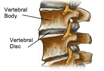
The discs of the spine sit between the vertebral bodies and act as shock absorbers for the spine. In the lumbar spine the outer part of the disc is made up of thick fibrous rings called the annulus fibrosus and the inner part is a soft jelly like substance called the nucleus pulposus. The discs in the spine also move to allow flexibility and movement of the spine, particularly in the lumbar spine where they are the largest.

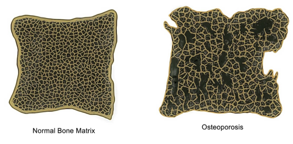
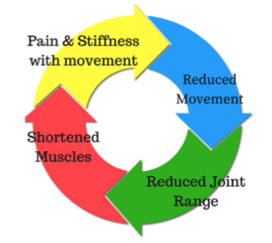
As we age, our discs begin to dehydrate, losing height, elasticity, and flexibility. The result is a spine that can feel stiffer, less flexible and a bit sore with certain movements. While almost everyone will have some degree of degeneration into their later years, not everyone will experience symptoms.


What are the Symptoms?

The most commonly felt symptoms of disc degeneration are stiffness and pain in the lower back. The pain is usually felt over the spine where the disc degeneration has occurred, but also often radiates into the buttock region. In severe cases of disc degeneration, increased wear and tear can cause bony spurs to develop on the vertebral body. These spurs can cause muscle weakness, numbness and tingling sensations in the leg and foot if they place pressure on the nerves around the spine.

How does this happen?


Disc degeneration usually affects people over the age of 50, however symptoms can begin in your 30s. As we age, all of our tissues gradually lose collagen and elastin, in the spinal discs this process results in smaller, less flexible discs. Repetitive activities like bending on the waist to lift heavy objects and prolonged sitting also cause additional wear and tear of the discs and amplify the degenerative process.

How Can Physiotherapy Help?

Imaging such as MRI and XRAY can help determine the level of degeneration that has occurred with increasing accuracy, however in recent years research has shown that the level of degeneration that can be seen In imaging cannot reliably predict the amount of pain a person will experience. This can be confusing for patients and having scans that show large amounts of degeneration can be a distressing experience, regardless of symptoms, making patients feel helpless.

Physiotherapy can help patients return to their normal activities as soon as possible and regain optimal function for their individual presentation. In addition, physiotherapy helps patients to manage their pain by helping them understand their pathology and, strengthen trunk stabilizers to prevent recurrence of any injury.

None of the information in this article is a replacement for proper medical advice. Always see a medical professional for advice on your individual injury.

Tips For Helping Kids of Today Stay Active

Research shows that many children today are struggling to meet their daily-recommended targets for physical activity. We know that inactivity is a risk factor for a multitude of chronic diseases and many of the habits that shape our adult lives are set in childhood. Physical activity is important for a growing body as movement and weight bearing have a large impact on bone strength, muscle and tendon health. Here are some tips to make sure your child is staying as active as possible.


  1. Find an activity that suits your child’s personality and abilities.


Children who are very coordinated and excel in competition may find team sports both increase their self-esteem as well as keep them fit. For other children, being a part of a team can cause embarrassment and they may say they dislike exercise, when in fact what they dislike is feeling like a failure and letting down the team.


These children may prefer a sport where success measured by improving on their own performance, rather than being compared to other children. Surfing, yoga, martial arts, dancing or gymnastics may be activities that suit your child if competitive and team sports cause them to be discouraged.


  1. Do get injuries checked out by a professional and invest in proper rehabilitation.


While children do bounce back quickly from injuries, they also may have difficulty expressing themselves and their worries when they have pain. A niggling pain that won’t go away may cause your child to say “I don’t like sport” rather than mentioning that they are in pain.


Some children may retain worries that they will hurt themselves again because of a previous injury and avoid exercise because they don’t feel completely confident. Your physiotherapist can help to identify any issues that your child is having and help to resolve them.


  1. Set fun and challenging goals for them to complete during their daily routine.


As less children are walking and riding to school, try to find ways to fit extra activities into the day. Have a daily long jump competition in the back yard at the same time each day or have bed making time trials. Have a routine before bed of age appropriate exercises, such as star jumps, hopping, balancing and running on the spot. You can make this part of the night or morning ritual, just like brushing your teeth.


Talk to your physiotherapist for more tips on how to increase your child’s activity levels. Finding an activity that matches your child’s age level is key to keeping them engaged and active. None of the information in this blog is a replacement for proper medical advice. Always see a medical professional for advice on your individual injury.


Osteoporosis Facts

Osteoporosis is a condition characterised by very low bone mass or density. This is caused by the body either losing too much bone, not making enough or both. Osteoporotic bones become weak and fragile and can break from small forces that would normally be harmless.

In osteoporotic bones, as well as loss of bone density and mass, there may also be abnormal changes to the structure of the bone matrix, which further contributes to the bone weakness.

Osteoporosis is an extremely common bone disease and women are more affected than men. As it is a progressive disorder that worsens with age, while the disease process might begin earlier, the effects are usually only noticed and diagnosed in people who are 50 years and older.

What are the Signs and Symptoms?


Often called a silent disease, many people with osteoporosis will have no idea that they have the disease, as there are no obvious symptoms. In fact, sometimes the first sign that an individual has osteoporosis is when the first bone is broken. Along with fractures, which are the most serious signs of this disease, osteoporosis can cause the upper back to become excessively hunched (itself often a result of spinal wedge fractures) and there maybe widespread pain as bony tissue is increasingly unable to withstand normal forces.

Fractures are a serious problem, especially in the elderly population. Bone breaks due to osteoporosis occur most frequently in the wrist, spine or hip. When the spine is affected by osteoporosis, people may develop a hunched or stooped posture, which can itself lead to respiratory issues and places pressure on the internal organs. Osteoporosis can severely impact a person’s mobility and independence, which can have a huge impact on quality of life.


What Causes It?


As this is primarily a metabolic disorder, there are a variety of things that can cause osteoporosis if they either interfere with the body’s ability to either produce bone tissue or encourage excessive breakdown. This can be anything from gastrointestinal conditions that prevent absorption of calcium, lack of dietary calcium or low levels vitamin D, which is essential for absorption of calcium.

Certain medications may also cause bone loss especially if they are taken for a long time or in high doses. A good example is the long-term use of steroids. Although steroids are used to treat various conditions, it has been proven that steroids can cause bone loss and eventually, osteoporosis.

As bones respond to force and weight bearing by building more bone, having a sedentary lifestyle or doing activities with low impact can also lead to osteoporosis and this has been shown be an issue amongst professional swimmers and cyclists.

How Can Physiotherapy Help?


Physiotherapy can help you to improve your overall bone health, avoid or recover from fractures. Physiotherapy exercises can direct you to safely increase your weight bearing, which can help build bone mass. Balance training is also an important factor as this can reduce the risk of falls. Your physiotherapist can also educate you on how to adjust your lifestyle, at home or at work, to protect your bones and improve your posture.

None of the information in this blog is a replacement for proper medical advice. Always see a medical professional for advice on your individual condition.

Achilles Tendon Tear

What is it?


The Achilles tendon is a band of fibrous tissue located at the back of the ankle. Its main role is to connect the calf muscles to the heel of the foot. This tendon is the largest tendon in the body and when it tightens, as the calf muscles contract, it pulls the heel allowing you to stand on tiptoe or to point your foot.

Achilles tendon tears commonly occur in athletes, however, this injury can affect anyone and surprisingly, a complete tear is actually more common than a partial tear.

These tears are commonly located at the part of the tendon where there is poor blood flow approximately 6cm above its attachment to the heel. Since there is poor blood supply, this part of the tendon is both vulnerable to injury and slow to heal.

What are the Symptoms?


Primarily, an Achilles tendon tear will cause difficulty in activities such as walking, running and jumping. Other signs and symptoms of an Achilles tendon tear include:

  • A loud pop or snap is heard
  • Sudden and severe pain at the back of the calf or ankle
  • Feeling of having been kicked in the calf
  • There is a gap between the tendon and the heel (about 2 inches above the heel)
  • Swelling and stiffness followed by weakness and bruising
  • Difficulty walking particularly during push off
  • Standing on tiptoe may be impossible

What Causes It?


Anyone can tear their Achilles tendon if the tendon is subject to excessive force or overstretching, however there are some factors that can increase your risk of injury. The most common activities that cause this injury are running and jumping.


The Achilles tendon can thin and weaken both as we age and also if it is not used. As a result of this weakening, it becomes prone to injury like tear or rupture with less force or stretching required before an injury occurs. A tear of the Achilles is often observed in people with pre-existing Achilles tendinitis. Other factors such as certain medications including antibiotics and steroids and some illnesses like diabetes and arthritis can also result in weakness of the tendon, increasing injury risk. Being obese is also a risk factor as excess weight puts additional strain on the tendon.

How Can Physiotherapy Help?


Treatment for Achilles tendon tear will depend on the patient’s age, how severe the injury is and the patient’s activity level. For young people especially athletes, they opt to have surgery while older people choose conservative treatments including physiotherapy.

Physiotherapy treatment for an Achilles tendon tear will involve exercises to strengthen the calf muscles and the Achilles tendon and exercises for stability. Many people are able to return to their normal activities within 4 to 6 months. Functional rehabilitation is also part of the program as it focuses on how you coordinate your body and how to move it. The aim of functional rehabilitation is to help you return to your highest level of performance.

None of the information in this blog is a replacement for proper medical advice. Always see a medical professional for advice on your individual injury

MCL Injuries

WHAT IS IT?

Your knee moves freely backwards and forwards; however the thought of it moving from side to side probably makes you cringe. This is because the knee joint has sturdy ligaments either side of it that prevent sideways movement and we instinctively know that a lot of force would be required to shift it in this direction.

The ligaments on either side of the knee are called the Medial Collateral Ligament (found on the inside the knee) and the Lateral Collateral Ligament (found on the outside the knee) and they each work to provide stability and restrict the knee’s movement into a sideways direction.


HOW DOES THIS INJURY OCCUR?


The typical mechanism for this injury is a force that drives the lower leg sideways away from the upper leg. This can occur from an awkward landing from a height, or when twisting with a foot fixed on the ground or from an external force hitting the outside of the knee, such as with a rugby tackle.


WHAT ARE THE SYMPTOMS?

MCL tears have quite a distinctive set of symptoms, with pain and swelling noticed quite specifically to the inside of the knee. The severity of the pain and swelling will be related to the number of ligament fibres damaged. Larger tears will also make the knee feel unstable or loose.

To classify the severity of the injury and help to guide treatment, a grading system is used. With grade 1 indicating that a few ligament fibres have been torn and grade 3 used for a complete tear of the ligament with associated joint laxity. Very severe MCL tears often also involve injury to the medial meniscus and ACL and can require surgical repair. However, most MCL sprains can be managed well with physiotherapy. Grade 1 and 2 MCL sprains take between 2-8 weeks to fully heal and a complete rehabilitation program is strongly recommended to prevent future injury.

HOW CAN PHYSIOTHERAPY HELP?

In the early stages of the injury, treatment is focused on pain and swelling management, while allowing the body to start the healing process through inflammation. This is best managed thought the R.I.C.E. principles (Rest, Ice, Compression and Elevation).

Following any injury, it is natural for muscles to waste a little and the damaged tissues to lose what we call proprioception, the ability to sense their own position in space. This loss of muscle strength and proprioception can contribute to further injury if not restored with a proper rehabilitation program.

Physiotherapy also aims to restore movement to the joint and support the ligament while healing to ensure that it is strong and healthy, and the scar tissue forms in an organized fashion, which makes the new ligament as strong as it can be and protects against future tears.


None of the information in this blog is a replacement for proper medical advice. Always see a medical professional for advice on your individual injury.

The Hidden Risks Of A Sedentary Lifestyle

By now it should come as no surprise that prolonged periods of inactivity are bad for your health. It seems that the science is in, and the bad news is that long periods of sitting or inactivity is a risk factor for many diseases, independent of other factors such as obesity.


What does this mean?

This means that if you are hitting the gym for an hour a day, but are sitting down for long periods during the rest of the day without moving, you may not be avoiding the health risks that come with sitting.

So why is sitting so bad?

There is evidence that shows that when sitting for long periods, our bodies show unhealthy changes in blood pressure and blood sugar levels that over long periods of time can lead to increased risk of diabetes, dementia and obesity. How much sitting is considered too much and how long it takes for these changes to take place is up for debate. What we do know is that regularly breaking up your periods of sitting is a healthy lifestyle choice you can make for yourself in the same way that drinking water, regular cardio exercise and eating vegetables are.

The insidiousness of the issue is that, it’s likely that you are sitting much more than you used to without even realising it. Car commutes to work are getting longer, working days are often spent at a desk and with the rise of online T.V, you might be guilty of three or more hours stuck in one spot catching up with your favourite characters before you know it.


I have an office job, what can I do?

The first step to changing this habit is to start being aware of exactly how long you are sitting for an uninterrupted period of more than 20 minutes. Once you start noticing your daily patterns you can identify moments where changes can be made. We’ve heard of incidental exercise, but we’ll call this ‘incidental standing’. Consider standing on the train instead of sitting or standing when you have a document to read.

Here are a few other tips to get you moving;

  • Set an alarm to go off every half an hour that reminds you to get up and get a drink. (This might also help to keep you hydrated)
  • Try out a standing desk.
  • Stand up to take phone calls or get up to go ask a colleague a question instead of calling them.
  • Take a walking lunch break.

Speak to your us for more ideas tailored to suit your individual lifestyle.

AC Joint Sprains (Separated shoulder)

WHAT IS IT?

The AC (Acromio-clavicular) joint is a thick fibrous joint that connects the top of the shoulder blade to the outer end of the collarbone. The joint is required to be strong and supportive and is the primary way in which weight bearing forces are transferred from the upper limb to the rest of the skeleton. The joint is connected by three strong ligaments, the Acromioclavicular, Corococlavicular and Corocoacromial ligaments.


HOW DOES THIS INJURY OCCUR?


The primary mechanism that will cause this joint and its ligaments to be injured is a force that separates the shoulder away from the collarbone, usually in a downwards direction. This can occur from a fall into the ground where the top of the shoulder hits the ground first, a rugby tackle or a fall onto an outstretched hand. As with all injuries, there are many variations in severity and a grading system has been developed to classify AC joint injuries.

WHAT ARE THE SYMPTOMS?

After an AC joint injury there is usually immediate pain on the top of the shoulder, swelling and bruising. There is often loss of movement of the shoulder, and pain from putting weight through the arm or carrying heavy objects. In severe cases there is a visible lump on top of the shoulder, known as a ‘step deformity’, which is where an obvious difference in height can be seen between the top of the shoulder and the collar bone. There is frequently pain felt when reaching across the body, as when putting on a seatbelt.

To confirm the diagnosis, your physiotherapist can perform some clinical tests and an X-ray can help to grade the severity of the injury. The classification that would be given to you by your physiotherapist or doctor help to determine the optimal course of action for each injury based in current research and available techniques.

There are different classification systems, some use four grades and other six. Injuries with a smaller number of ligament fibres being torn are given a lower grade classification, going upwards as further damage is incurred. Injuries classified as higher grades will require surgical repair.

HOW CAN PHYSIOTHERAPY HELP?

The role of physiotherapy in this case is to ensure the joint is supported and given a chance to heal naturally, while maintaining the strength and normal movement of the shoulder girdle. This is done initially by providing support to the joint. You may need to have your arm supported in a sling or brace for some of this time and your physiotherapist can show you some taping techniques to add support.

Most AC joint sprains take six weeks to fully heal, although many patients report shoulder problems in future years. For this reason a comprehensive rehabilitation program is very important. More severe sprains are often treated with surgery to stabilise the joint and treat any possible fractures. Surgical repair will also require a proper rehabilitation program.

None of the information in this blog is a replacement for proper medical advice. Always see a medical professional for advice on your individual injury.

Understanding Joint Stiffness

While pain and stiffness often go together, joint stiffness can occur on its own. Joint stiffness can limit your ability to perform usual tasks, for example turning your neck to check behind you while driving. Stiffness can also be a warning sign that part of the body is vulnerable to future injury. There are many different causes of stiffness and we will explore a few of the reasons why you might not be feeling as flexible as normal.


  1. Disuse and lack of movement.

Our bodies are made to move. When we are not regularly moving them through their full range, they can begin to feel ‘tight’. This can be caused by a combination of the capsule that surrounds the joint tightening up and the muscles that surround the joint shortening and losing flexibility. Stiff and tight muscles can cause you to feel as though your joints are stiff, even if it is only the muscle length that is restricting the movement. Joint mobilizations, manipulation and muscle stretches/massage can have a significant effect in improving the symptoms.

The most important way to maintain full movement is to regularly move joints through their full range, which also helps to keep muscles and joints healthy. Your physiotherapist can advise you on how to best approach this with a targeted set of exercises.

  1. Osteoarthritis (OA):

OA is a degenerative disease, characterised by a breakdown of joint surface cartilage and the growth of bony osteophytes around areas of stress. A person with OA will usually feel stiff for around 15-20 minutes after being still. Physiotherapy programs to strengthen the muscles surrounding the joints, so as to help absorb weight-bearing forces, has been shown to have positive results on OA symptoms. While OA is increasingly common as we age, it is thought that the primary cause is abnormal load and stress to joint surfaces and not simply aging itself. As the joint space between two joint surfaces becomes uneven, joints affected by OA can feel stiff or even ‘blocked’.

  1. Inflammatory Related Stiffness.

The inflammatory process is characterized by swelling and pain around a specific area. Usually this is a response to damage by the body. As an area swells, this will allow less space for movement and a sensation of stiffness, as anyone who has had sprained an ankle can attest to. Acute inflammation will cause swelling that increases over 24-48 hours and subsides gradually. Autoimmune disorders can cause the body to mistakenly have an inflammatory reaction where there has been no injury, with resulting pain and stiffness. Rheumatoid arthritis and ankylosing spondylitis are two examples of such disorders.

Stiffness caused by inflammatory disorders is characterized by feeling of stiffness after rest, particularly in the morning that can take longer than 30 minutes to subside. Inflammatory disorders unrelated to injuries are complex in cause and require collaboration with medical teams for best treatment outcomes. Acute injuries are best managed by following RICE protocols (Rest, Ice, Compression, Elevation). Speak to your physiotherapist for more information regarding a specific condition.


LCL Tears

The knee is one of the largest joints in the body and has only one plane of movement. This means it bends and straightens but does not twist (much) or move from side to side. To keep the knee from moving in other directions, the knee is supported by many strong ligaments, with two of these being found on either side of the knee. The inside ligament is the ‘Medial Collateral Ligament’ (MCL) and the outside one is the ‘Lateral Collateral Ligament’ (LCL). The primary role of the LCL is to prevent the lower leg from moving too far towards the midline in relation to the upper leg.  Both the LCL and MCL are extremely strong ligaments and provide lots of support to the knee during movement however, they are still vulnerable to injury.


How do tears happen?

The LCL is injured less often than the MCL, however tears do still occur. The most common way the ligament is damaged is through a force causing the knee to move inwards in relation to the upper leg, or a twisting of the knee. This can be seen in sports that involve changing directions or with a direct force, such as a rugby tackle. This injury can also occur from a simple fall and as with all sporting injuries, it is not only athletes who can be affected, anyone can tear their LCL in the right circumstances.


What are the symptoms?

Following an injury to the LCL, common signs and symptoms are a ‘popping’ sound at the time of injury, immediate pain with weight bearing and swelling and a feeling instability. The severity of the injury will impact how much each of these symptoms are felt and LCL tears are classified as either Grade I, II or III, which helps to direct treatment. A grade I tear is where a few fibers of the ligament are stretched and damaged, a grade II is where this a partial rupture of the ligament with some instability of the knee and Grade III is a complete tear.


How are LCL tears diagnosed?

Your physiotherapist is able to perform clinical tests to evaluate if there is any instability of the knee from an LCL tear. An MRI can confirm this diagnosis and an X-ray may be required to rule out any associated fracture. It is possible for nerve damage to occur at the same time as an LCL Tear, which will result in weakness and loss of sensation in the lower leg. Severe injuries are more likely to involve injury to other parts of the knee and your physiotherapist will make a full evaluation of all your injuries on assessment.


Most LCL tears are managed well with just physiotherapy and support of the joint, however severe tears and associated nerve damage may require surgery. Your physiotherapist and medical team will work together to help determine the best course of action for each individual injury.


How can physiotherapy help?

For tears that don’t require surgery, your therapist will advise you on how to best support and protect the injured joint. In the first 48 hours, RICE protocol (Rest, ice, compression, and elevation) is applied to reduce any pain, swelling, and inflammation. Following this period, you will be advised on how best to mobilise the joint whilst preventing any further damage. Return to sport will be dictated by healing times with a full recovery expected by 6-12 weeks.

Following ligament damage, balance, strength, and proprioception are often impacted and your physiotherapist will develop a program to address this, which is an important part of preventing further injury. Tears that are repaired surgically will require a longer program of rehabilitation and close liaison with the medical team.

None of the information in this blog is a replacement for proper medical advice. Always see a medical professional for advice on your individual injury.

How Does Diabetes Affect Healing Times?

It can be surprising to many people that one of the questions their physiotherapist will ask them when assessing an injury is ‘do you have diabetes’. This may seem more like an issue for your doctor than your physiotherapist!

The reason why your therapist is asking is that diabetes can actually have quite a large effect on healing times of body tissues. At times, injuries can take up to twice as long to heal properly in patients with diabetes and your physiotherapist will need to update their training and rehabilitation programs to factor this in.

How does this happen?

From what most people know about diabetes, it seems strange that it would affect healing times. However, the more you understand about the processes that cause diabetes the more sense it makes.

It all comes down to blood flow. Our veins and arteries are made of flexible and elastic tissues that expand and contract when necessary to allow the optimum amount of blood flow to an area. Sometimes is it better for tissues to have less blood flow and other times they require more. This flexibility of the blood vessels is essential for controlling and modulating the amount of blood to an area at any given time.

When someone has diabetes, they have an excess of glucose in their blood. This occurs because the body is unable to regulate insulin, which is used to break down glucose and

provide the body with energy. This can be due to an autoimmune disorder that affects the cells that make insulin (Type I) or insulin resistance due to dietary choices (Type II). Over time, this excess glucose sticks to the blood vessel walls and they can become harder, losing their elasticity and ability to change size rapidly. Primarily this will affect the ability of the blood vessel to expand reducing the amount of blood flow available to the tissues.

Diabetes can also affect the health of the nerves in they body, particularly in the hands and feet. This can result in poor sensation, which means that the person may not realize when the injury is being further injured.

What does this mean for recovery times?

While not everyone with diabetes will have this issue, it is something that needs to be made known to your physiotherapist so they can be aware of the possibility. These changes are more likely to occur after having diabetes for a long period of time and if it is poorly managed.

Ask us for more information on how diabetes may be affecting your recovery and for tips to ensure the best outcomes possible.