Hip Pain and Labral Tears

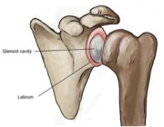
What are Labral Tears?

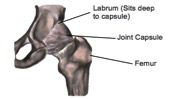
If you are experiencing pain in the front of your hip along with clicking, locking or catching of your hip joint you may have underlying labral damage. The acetabular labrum is a fibrous rim of cartilage that covers and seals your hip socket. This lining of cartilage provides stability for the thigh bone (femur) inside of the hip socket (acetabulum).

The labrum seals the hip socket, providing a suitable appropriate rotational axis for the thigh bone in the hip socket as well as helping to maintain the nutritional fluid within the joint that is important to maintain joint health.

How do they happen?

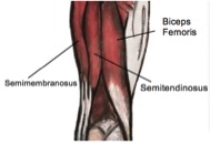
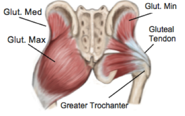
Labral tears can occur from an injury such as a twist or slip, or damage can occur from repetitive stresses. Anatomical changes in normal hip movement, which may also be associated with neuromuscular imbalance most commonly causing labral tears, are repetitive movements where there is decreased joint clearance between the femur and the acetabulum. For example athletes such as gymnasts and ballet dancers who have to repeatedly pivot or flex their hip are more likely to damage their labrum than those who do not. Over time this repetitive impingement of the hip joint can cause the labrum to tear and damage to the labrum if not managed properly may lead to early degenerative arthritis.

What are the symptoms?


Pain in the front of the hip, groin, side of the hip or buttock often described as deep are symptoms of labral damage as well as clicking, locking, catching or giving away of the hip. Prolonged sitting, standing, walking or pivoting can cause pain for someone with a labral tear and this may cause a limp when walking. Other signs and symptoms include joint stiffness or a feeling of instability in your hip.

How are they Diagnosed?

Diagnosis is not always possible to confirm in the clinic, however, magnetic resonance arthrography (MRA) has been found to be very accurate in diagnosing labral tears. An MRA is when a dye is injected into the hip joint before the hip joint is scanned and specialist photographs of the joint are taken. Arthroscopies are another option but as they are more invasive they are often not the first port of call.

It is advised to speak to your physiotherapist about your symptoms who will gather a thorough history of your problem and may undergo a series of tests as part of the physical examination. A physiotherapist can inform you if they think your symptoms are coming from labral damage or if they suspect a different problem.

How can Physiotherapy help?


If your physiotherapist does suspect you have a labral tear, different treatment plans are available. Physiotherapy management may include a stretching and strengthening program to correct any neuromuscular imbalance; movement re-education and a variety of manual techniques that can be performed by your physiotherapist may reduce or abolish your symptoms. Other options are available or may be used in conjunction to physiotherapy including pharmaceutical medicine, corticosteroid injection and surgery and you should speak to your physiotherapist and doctor about this.

None of the information in this blog is a replacement for proper medical advice. Always see a medical professional for advice on your individual injury.

Understanding Hamstring Tears

Let’s get clued up on hamstring strains; why you may have one, what you can do to help and how to prevent a future injury. The hamstrings are a group of three muscles; the biceps femoris, semimembranosus and semitendinosus. You can feel these muscles if you place your hands on your sitting bones where the muscles originate and slide your hands down the back of your legs. The main action of these muscles is to bend your knee, take your leg out behind you and to assist rotation of your knee, especially when performing accelerating and decelerating actions.

A strain/pull/tear is when the muscle fibres are overstretched. Injuries are frequently felt as a short sharp pain in the back of your thigh whilst exercising. A hamstring strain will typically happen when running just before your foot hits the ground. At this point, the hamstrings are working eccentrically to control the forward motion of the two lower leg bones, your tibia and fibula. Pain is often the most debilitating symptom affecting your ability to continue exercising and may cause a limp. Other symptoms include swelling, bruising, muscle spasm and reduced movement at your knee.

Strains can be categorised into 3 different grades. 1 being the mildest with a small number of fibres being torn to grade 3 being the most severe which can be a complete muscle rupture. The good news is muscles have a fantastic blood supply and should heal within 3-12 weeks depending on the degree of injury. However, the flexible skeletal muscle fibres, which your muscles are made up of, are replaced with much more inflexible tough scar tissue, which is where physio’s come in. Specific rehabilitation such as specialist stretching, strengthening, taping and soft tissue techniques can dramatically influence how muscle fibres are restructured reducing the amount of scar tissue speeding up the healing process helping you return to sport quicker. With any soft tissue injury, R.I.C.E (rest, ice, compression, elevation) should always be your first response.

A physio will be able to perform a thorough assessment and educate you on why you sustained a hamstring strain in the first place. Common factors that can predispose you to hamstring strains are not warming up or cooling down properly, tight hamstrings or hip flexors, weak hamstrings or gluteal (butt) muscles, training at a high intensity without adequate training or altered biomechanics.

Runners often have short, weak hamstrings, tight hamstrings will restrict the length of your strides when running meaning you have to work harder to cover the same distance as you would with adequately lengthened hamstrings. Chronically tight hamstrings can cause not only hamstring strains but can contribute to back pain, knee pain and leg length discrepancies. So even if you have never stretched before it may be a good time to start stretching!

So to prevent yourself pulling a hammy make sure you warm up and cool down properly including effective stretching of not just your hamstrings but hip flexors, quadriceps and calf muscles, do sport specific strength and conditioning and avoid sudden increases in intensity of exercise. On your next visit why not ask your physio and find out how healthy your hamstrings are

Gluteal Tendinopathy

What is Gluteal Tendinopathy?

When tendons are repeatedly placed under more tension than they can deal with, they can have a failed healing response. This can cause changes to the structure of the tendon and is known as a tendinopathy. When this occurs in the tendons of the gluteal muscles it is referred to as gluteal tendinopathy.

The gluteal muscles are three large muscles located at the back of the pelvis that provide most of the muscle bulk of buttock region. These muscles work together to keep your pelvis level when standing and are responsible for many movements of the hip. They play an important role in standing, walking and running.

The two deepest gluteal muscles, gluteus medius and gluteus minimus, attach from the center of the pelvis (the sacrum) and insert into the bony outer region of the upper thigh, called the greater trochanter via the gluteal tendons.

What causes tendons to develop tendinopathy?

Tendons, like muscles, skin and bones are living tissues and their strength and elasticity is influenced by a variety of factors, including hormones, age, how often and how much they are used. Rapid changes in activity levels or simply performing the same tasks too often can place a tendon under more stress than it can tolerate and it begins to break town.

Recently it has been shown that tendon health is also negatively affected by compressive forces, which can occur from blunt trauma or even habits such as crossing the legs, or sleeping on your side on a hard mattress.


What are the symptoms of Gluteal Tendinopathy?

When gluteal tendons are affected by tendinopathy, a typical pattern of sharp pain at the outside of the hip with specific movements is present. The pain is usually worse with walking, going up and down stairs and running. The pain can become quite severe, and eventually can impact day-to-day activities.


How can physiotherapy help?

A thorough assessment is required for an accurate diagnosis and once gluteal tendinopathy is confirmed, your physiotherapist will be able to identify which factors have contributed to your condition and help to address these. It has been shown that specific loading exercises and muscular retraining can stimulate the tendon to heal and remodel the collagen fibres into a more organized pattern again. Your physiotherapist can investigate any postural habits or activities are contributing and address these as required.

None of the information in this blog is a replacement for proper medical advice. Always see a medical professional for advice on your individual injury.

10 Facts About Tendons

Tendons are found all over the body and while you may know a little about them, you might be surprised to learn a few of these facts.

  1. Tendons can be found at the ends of muscles. Tendons are simply connective tissues that attach muscles to bone and help them move our joints when they contract.
  1. Tendons come in many shapes and sizes. While the most recognizable shape is the long thin kind (such as the Achilles tendon), they can also be flat and thin or very thick, depending on the shape of the muscle and attachment of the bone. A thin flat tendon is also known by the name aponeurosis.
  1. Tendons are able to act like elastic bands, they can stretch and bounce back into shape. Like elastic bands, if too much force is applied they can stretch or tear.
  1. Unlike elastic bands, tendons are living tissue and their properties are affected by many different factors. Seemingly unrelated things such as hormonal changes, autoimmune disorders and nutrition can all affect a tendon’s ability to withstand load.
  1. Tendons don’t only attach muscles to bone, they can attach to other structures as well such as the eyeball.
  1. Tendons can tear however; more often they are injured through overuse. Healing of tendons can be quite slow as they have less blood supply than other tissues of the body, such as muscles.
  1. Tendons are mostly made of organized collagen fibres. Areas of tendon degeneration have been shown to have collagen fibres that are disorganised, with this area having less strength and elasticity.

8. The Achilles tendon is the strongest tendon in the body. This connects the large calf muscles to the back of the heel to point the ankle away from the body. Most tendons are simply named for the muscle they attach to, however the Achilles has it’s own name, named for the mythical Greek character whose heel was his only point of weakness.

  1. The smallest tendon is located in the inner ear, attaching to the smallest muscle in the body.

10. Tendons and muscles work together to move your joints and are called a contractile unit.

Acute Wry Neck

Have you ever woken up with an inexplicably stiff and painful neck that will only turn to one side? You might have been suffering from acute wry neck, a painful condition following a typical pattern of symptoms. In the clinic, wry neck is classified as one of two different types – Facet or Discogenic wry neck. These have similar presentations, yet are caused by slightly different things and require different treatment.

Facet Wry Neck:


Facet joints are found on either side of each vertebrae of the spine and allow for controlled rotation and side bending. An awkward movement of the neck can cause a part of the joint capsule to tear or get caught in the joint, making it feel locked. The muscles around the area can also be come tight and spasm, which contributes to the problem. The pain is usually sharp and can be pin pointed quite accurately to the part of the neck causing the problem and the pain rarely travels down into the arm. It is usually possible to find a resting position where the pain goes away completely, only having pain when turning in certain directions.

The good news about Facet Wry Neck is that your physiotherapist is usually able to help you ‘unlock’ the neck quite quickly with gentle mobilisations and get you back into action. Most of the time a full recovery can be expected within a week.

Discogenic Wry Neck:


Each of the vertebrae of the neck are separated by fibrous disc that are filled with a gelatinous center. These discs provide support, flexibility and shock absorption. Under pressure, these discs may bulge or tear and the resulting swelling can cause pain and muscle spasm in the surrounding area. It is important to note that in an area as sensitive as the neck, a small amount of damage can result in a large amount of pain.

The development of discogenic wry neck is usually due to a combination of factors, including neck stiffness, poor posture and biomechanics than can contribute to the disc being vulnerable prior to injury. In this case it can be difficult to find a comfortable position and it is more likely for the pain to travel into the arm and may even be associated with pins and needles.

Treatment of discogenic wry neck will be focused on reducing pain and muscle spasm with massage, taping, heat and postural education. Further treatment aims to reduce any stress that is being placed on the disc, mobilise any stiff spinal segments and correct any muscle imbalances. While the initial symptoms may settle down quite quickly, it can take up to six weeks to fully recover from discogenic wry neck. In this condition it is also important to address all the factors that may cause a recurrence of the issue.

wry neck

Labral Tears Of The Shoulder

What is the Labrum of the shoulder?

The shoulder is a remarkably mobile joint, however this flexibility comes with the cost of less stability. The glenohumeral joint, where the upper arm meets with the shoulder blade is a ball and socket type joint. The surface area of the ‘socket’ part of the joint (the glenoid fossa) is actually much smaller than the ball part of the joint (the head of the humerus). A fibro-cartilaginous ring called a labrum, surrounds the edge of the glenoid fossa which acts to increase both the depth and width of the fossa.

This labrum provides increased stability and is also the attachment for a part of the biceps muscle via a long tendon. The labrum is able to provide flexibility and stability that a larger glenoid fossa might not be able to, however being a soft structure it is prone to tearing which can be problematic

What causes the labrum to tear?

The most common way the labrum is torn is through a fall onto an outstretched arm or through repetitive overheard activities such as throwing or painting as the repeated stress on the labrum can cause it to weaken and tear.

Suspected labral tears can be diagnosed in clinic by your physiotherapist through a series of tests, however an MRI is required to fully confirm the presence of a labral tear. Labral tears are classified into different grades, which are determined by their location and severity. This grading is used as a guide to help determine the correct treatment.


What are the symptoms of a labral tear?

A labral tear is often associated with other injuries, such as rotator cuff tear, which can make the clinical picture a little confusing. Commonly there will be pain in the shoulder that is difficult to pinpoint and the pain will be aggravated by overhead and behind the back activities. Severe labral tears can lead to instability and can also be related to dislocations of the shoulder.


How Can Physiotherapy Help?

The severity and grade of the labral tear will guide treatment. Smaller tears can be treated with physiotherapy that is aimed at increasing strength and control of the shoulder. Other tears may require surgical repair after which physiotherapy is an important part of treatment to rehabilitate the shoulder.

labrum tears shoulder

None of the information in this blog is a replacement for proper medical advice. Always see a medical professional for advice on your individual injury.

A Global Campaign To Improve Balance

Keeping your balance is a complicated process. Although you might not realize it, your body is constantly making calculations and tiny postural readjustments to stay upright.


There are many parts of the body that work together to maintain balance, and this means that if one body system, for example vision, begins to fail, the other systems are able to compensate. The body is so good at doing this that you might not even notice your balance is deteriorating unless put under more difficult conditions.

Poor balance is associated with falls, longer hospitalization, permanent injury and even fatality. The good news is that balance can usually be improved. Your physiotherapist is an expert in balance and understands how to identify which aspects of your balance needs addressing and how to improve them.

Recently a global physiotherapy awareness campaign was launched called “One Leg Physio” which issues a challenge to people to reach their highest level of balance by working through a progressive list of challenges called the ‘difficult dozen’.

The challenge is to make your way safely through as many the difficult dozen as you can and hold for at least 34 seconds.

You can participate in this global challenge yourself by going to the website, www.oneleg.physio and trying out the difficult dozen for yourself. Make sure you do so safely and protect yourself if there is any risk you might fall.

To help spread awareness, the campaign asks you to post pictures of your most successful attempt and post to social media with the hashtag #onelegphysio and tag two friends to challenge them. You can also compare your best times with those of people from all around the world. Don’t forget to tag the clinic as well!

If you want to improve your balance, talk to your physiotherapist about developing a specialized program. They can do a thorough assessment to work out the most appropriate exercises to make the most improvements to your overall balance. This can be an important factor in preventing falls and injury, no matter your fitness level or age.

Sever’s Disease

What is Sever’s Disease?


Sever’s disease is a condition that causes pain in the heel of children and adolescents. While it can be quite painful, it is a self-limiting condition that doesn’t usually cause any long-term problems. Sever’s disease is quite common and is actually the leading cause of heel pain in children and young adolescents.

Also known as calcaneal apophysitis, Sever’s disease is an inflammation of the growth plate of the heel. A growth plate is the area of bone where bone is produced in the growing skeleton, and is often weaker than other bony areas. Repeated or excessive stress on this area can cause it to become and inflamed and painful.

sever's

What are the symptoms?


Children might complain of heel pain that is worse when walking, running or jumping. This may be present following an increase in activity or after a growth spurt. Pain may also be felt when walking on heels.

Why does this happen?


The Achilles tendon attaches to the back of the heel, just next to the growth plate and puts force through this area during gait. If this force becomes too great, this growth plate can become irritated, starting a painful inflammatory process.

While increased activity is definitely a predisposing factor, other things may also contribute, such as; reduced movement of the ankle, abnormal foot movements when walking and tight calf muscles. It is also possible that Sever’s disease will appear with no apparent cause.

What is the treatment?


While this is a self-limiting disorder that will go away on it’s own as the skeletal system completes growth, it can be quite painful and this may impact your child’s activity levels and gait pattern.

To prevent any long-term issues from adaptations or changes in activity levels, your physiotherapist can work with your child to find solutions that allow maximum movement with minimum pain. Most of the time, simple education and relative rest or ceasing of aggravating activities are effective, however recent studies have found that properly fitted orthotics can help reduce pain while maintaining activity. Physiotherapists can also address any factors, which may have caused excess stress in this region, such as abnormally tight muscles or poor gait patterns. They can also advise of pain relieving treatments and alternative forms of exercise if necessary. Speak to your physiotherapist for more information.

None of the information in this blog is a replacement for proper medical advice. Always see a medical professional for advice on your individual injury.

FOCUS ON SHIN SPLINTS

What is a stress fracture?

A stress fracture is a microscopic fracture of the bone that is so small that it often cannot be picked up on X-ray. If left untreated, a stress fracture can cause significant disability and develop into a full fracture, possibly even requiring surgery.

The majority of stress fractures occur in the lower limb, being particularly common in the hip, shins and foot at points where the most force passes through when weight bearing. Most stress fractures are overuse injuries and are common in long distance runners.

 Shin Splints

What are the symptoms?


As with many overuse injuries, the pain of a stress fracture starts gradually, beginning with pain during or after activity or sometimes the morning after. If activity continues without modification, the pain will gradually increase. Eventually most people are unable to maintain their usual activity level. Stress fractures are very are common in runners and military personnel who are required to march for long periods. A stress fracture will be more likely to occur in a person who has weaker bone strength, such as someone with osteoporosis, which is itself affected by many factors such as adequate calcium intake, vitamin D deficiency and a history of inactivity.

How are stress fractures treated and how long will it take to get better?


Stress fractures can easily be mistaken for other conditions such as shin splints. As the fracture is often too small to show up on X-ray, definitive diagnosis can be made using MRI or bone scan.

After diagnosis, the most important part of treatment will be resting the area to allow the bone to heal before resuming activity. Stress fractures usually need at least 6 weeks to recover fully. Some areas of the body have poor blood supply, which makes healing more complicated. For example, stress fractures of the navicular bone of the foot may need to be kept still and placed in a boot or cast for a period of time to heal properly.

Other aspects of treatment will involve correcting any factors that contributed to the original injury. There is some evidence that unsupportive footwear is a risk factor, along with poor biomechanics and weak muscles that provide inadequate support to the skeletal system during activity. Speak to your physiotherapist if you suspect you may have a stress fracture or simply want to know more.

None of the information in this blog is a replacement for proper medical advice. Always see a medical professional for advice on your individual injury.

Healthy Lifestyle Changes To Make This New Year

Most of us start the New Year with some ideas of how we will change our lives for the better. Here are some tips for a better lifestyle and healthier outlook for the coming year.


Be proactive about your health.Happy New Year


Take time to listen to the advice of health care professionals and seek treatment for niggling issues before they become bigger.


Aim to be a little bit more active every week.


You can include any kind of activity, just try to move a little bit more each week. There is nothing more disheartening than starting a new hobby and increasing your program too quickly only to develop an injury that sets you back. Pace yourself with the view of realistically maintaining your new activity.


Take the advice of health professionals.


Physiotherapists know only too well that their advice is not always taken seriously. Unfortunately, some conditions do take hard work and time to resolve. Quick fixes only exist for relatively few conditions and the reality is that problems that develop over a few months don’t usually resolve overnight. Follow your physio’s advice for best outcomes. (And remember to do your home exercise program!)


Use exercise as a way to improve social and mental wellbeing.


While any amount of exercise will have a positive effect on your mental health, you can use a new hobby to expand your social circle and even improve self-esteem as you find yourself mastering a new skill. Try something a little different like rock climbing or skateboarding.


Pay attention to your diet.


While dietary fads are questionable in their effectiveness, there is no harm in monitoring what you are eating just a little more closely in the New Year. Remember to balance your meals with enough carbohydrates, protein, vitamins and minerals.


Swap sugary drinks for water.


Adequate hydration is an important part of keeping your body functioning well. Sugary drinks can also add a deceptive amount of calories to your daily intake.