Strains vs. Sprains

Strains and sprains are words that are used almost interchangeably when describing injuries, however they each actually have quite distinct meanings. The most straightforward explanation is that a “strain” refers to a tear in a muscle or tendon, while a “sprain” refers to a tear in ligament fibers. Below we briefly describe what that means and how we treat sprains and strains differently.


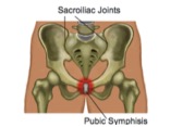
Ligaments are fibrous tissues that connect and hold bones to other bones. These are very strong parts of your anatomy and, depending on the joint, provide large amounts of support and stability to the body.


Some ligaments are so strong, for example in the shoulder, that sometimes a bone will break before the ligament will tear. When ligament fibers do tear, the nearby joint can actually feel unstable.


Ligament tears will usually swell up and have a normal inflammatory reaction. Pain will be worse with movement or if the ligament is placed under more stress. Occasionally, if a ligament has torn all the way through, there is much less pain than with a less severe injury.


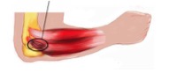
Your physiotherapist is able to grade the severity of a ligament sprain, which will help guide treatment and expected recovery times. Muscle strains are easy to confuse with ligament sprains, however there are a few telltale differences. Following a muscle tear, it is more likely that you’ll feel weakness rather than instability. The pain will also be isolated over the muscle or tendon body. An injury to a ligament will be tender over the site of the ligament and special tests can be done to show a laxity due to the injury.


Treatment is also slightly different as sprains will need more support and will sometimes even need to be braced, whereas muscle strains will benefit from gentle movements earlier. In both cases, following the basic principles of rest, ice, compression and elevation is great advice in the early stages of any injury. Heat is not recommended until at least two days after the injury.


It is important to seek professional opinion when recovering from both a strain and a sprain. It is very easy to re-injure an area while it is healing if undertaking strenuous activity too early and without correct rehabilitation. Speak to your Matthew for more information.

Metatarslagia

What is Metatarsalgia?


Metatarsalgia is a medical term used to describe pain that occurs in the ball of the foot. This occurs due to rubbing of the long bones of the foot, called metatarsals, causing inflammation. Build-up of inflammation in the ball of the foot compresses the small nerves that run between the toes and along the foot. Inflammation and compression of these small nerves is painful.

The pain can involve one or more joints of the foot including the big toe, second toe and third toe. It is unlikely to include the fourth or fifth joints. On occasion, the pain may involve the entire foot, or larger parts of the foot.

The pain or symptoms may be made worse when bearing weight through the foot, such as walking or running. In severe cases, just standing or even wearing tight footwear can affect the foot.


How does it happen?


Metatarsalgia can be caused by a number of things. An abrupt change in posturing of the lower back or pelvis may alter the way the foot is loaded, and cause rubbing of the long bones. Altered loading of the foot may also be caused arthritic conditions that affect the knees, hips or pelvis and carrying extra weight.


Unavoidable factors, which may lead to the development of metatarsalgia, include increased age, and the physical shape of the foot and toe. As your body ages, the ligaments of the foot may loosen or weaken. Therefore, the bones of the foot move differently leading to altered positioning and loading of the foot.


Avoidable factors, which may lead to the development of metatarsalgia, include wearing inappropriately fitting footwear, and the type of exercise performed. Metatarsalgia is more highly associated with high impact-type exercise such as running or gymnastics.


What are the signs and symptoms of Metatarsalgia?


The signs and symptoms of metatarsalgia may vary from person to person. Generally, a complaint of pain is common. This may range from mild to severe, or may be described as burning or shooting pain in the foot or toes. Another common description is a tingling sensation in the foot or toes.


Typically, symptoms worsen when weight bearing and improve once weight is removed from the foot.


How can Physio help?


Your physio will determine the cause of the metatarsalgia pain and assist you with adapting your posture, or improving the loading pattern of your foot. You may be advised to unload the foot by reducing or stopping exercise, changing the shoe you wear, or strengthening certain muscles in your legs.


Electrotherapeutic modalities such as icing, ultrasound or interferential therapy may be of assistance to reduce pain and inflammation in the beginning stages of treatment as well.


The information in this newsletter is not a replacement for proper medical advice. Always see a medical professional for assessment of your individual condition.


Orthopaedic Surgery And Physiotherapy

Orthopaedic surgery is any surgery performed primarily on the musculoskeletal system of the body. Common orthopaedic surgeries you may have heard of are joint replacements and reconstructions.

Physiotherapists have a large role to play in the recovery of these kinds of surgeries especially in the more extensive operations, such as a total hip or knee replacement.

It is not unusual to wake up in the hospital a few hours post-op knee replacement, hip replacement or shoulder reconstruction to your hospital-based physio asking you to get out of bed for the first time. This may seem daunting and entirely too early, however there is very well researched evidence for why getting out of bed early is so important: your speedy return to normal health and wellbeing!

Hospital-based physiotherapy typically includes re-learning how to walk and making initial improvements on your range of movement and function of the affected joint. The physio will also help you maintain healthy respiratory function by checking your lungs regularly. Drugs administered during surgery change your breathing pattern, which can lead to complications in the days after surgery.

Following your in-patient stay, it is highly likely your surgeon will request for you to continue with outpatient physiotherapy either in an outpatient hospital-based setting or in a private-practice setting.

With your outpatient physio, your recovery will be measured and monitored, goals will be discussed and set, and you will be given an exercise program created to achieve those goals.

Physiotherapists use a variety of treatment methods to assist a post-operative orthopaedic surgery patients achieve their goals. These include soft tissue work, stretching, hydrotherapy, manual therapy and gym-based exercise.

Recovery from orthopaedic surgery can take up to 12-to-18 months to occur. During this time, it is not unusual to experience bouts of pain and swelling; however these episodes should reduce the further into rehabilitation you progress.

Many post-operative orthopaedic surgery patients can expect to return to a normal life following a period of rehabilitation guided by a physiotherapist. This includes return to some sport and regular exercise!

Note: Please seek professional advice prior to beginning a post-operative exercise program.

Osteitis Pubis

What is Osteitis Pubis?

Osteitis Pubis is a medical term used to describe sports-related groin pain. Osteitis means ‘bone inflammation’, while pubis refers to the specific bone that is affected: the pubic bone. Osteitis pubis is usually an overuse injury that can sometimes be triggered by a specific event. It is characterized by pain deep within the front of the pubic bone, caused by inflammation. The area of the pubic bone affected is specifically known as the ‘pubic symphisis’.

This type of injury is common in load-bearing athletes such as runners. Other people commonly affected include soccer players and footballers, due to their frequent kicking actions.


How does it happen?

Instability within the pelvic region is the primary cause of Osteitis Pubis, particularly if the instability occurs at the connection between the two sides of the pubic bones at the front of the body. The pelvis carries the weight of the upper body and is responsible for providing stability when walking, running and kicking. This means that the joint can easily become irritated and inflamed.

What are the signs and symptoms of Osteitis Pubis?                                                  

Osteitis pubis is aggravated by weight-bearing activities, with running and kicking being the two main culprits. Pain is usually experienced on one side, however both sides can be affected. The pain is usually located at the front of the pelvis, and may progress into the hip and groin area as it becomes more severe.

Sufferers of Osteitis Pubis tend to have a history of previous groin strain, as well as lower back pain. They may also have a history of a sports hernia in the hip area. As with most inflammatory conditions, the pain may be worse when in use, better when resting, and worse overnight into the morning.

 How can Physio help?

Your physio is able to help this condition in a number of ways and will hopefully get you back to your pre injury sporting level. During assessment, your physio will look at many different things to determine the cause of the condition. Muscle length, muscle strength and muscle control will all be assessed. Your posture in standing, walking and running will also be assessed to determine any irregularities.

Your physio will ask you to rest from sport for a period of time to allow some bony healing to occur. They will then progress you through a rehab program aimed at getting you back to sport.

This rehab program will retrain your muscles to stabilize the pelvis when walking, running and kicking. The muscles will also need to have relatively equal flexibility to help stabilize the pelvis. Your physio will give you specific exercises to target the strength and flexibility of these muscles. Finally, your physio will progress you to running or kicking, and allow you to gradually return to sport over a 3-to-6-month period of time.


The information in this blog is not a replacement for proper medical advice. Always see a medical professional for assessment of your individual condition. 

Recognising and Managing Ankle Fractures

Ankle fractures are surprisingly common injuries, both in athletes and the general population. Being able to quickly identify the signs and symptoms of a fracture is extremely important in ensuring quick and effective medical management.


Ankle fractures come in different shapes and forms. For example, a netballer with shin splints may experience a stress fracture, while a soccer player colliding with another player may experience a large break of the bone at the base of the ankle. In either case, bony tissue has been disrupted and must be treated accordingly.


Knowing the signs and symptoms of fracture


Understandably, swelling and bruising will occur when a bony injury to the ankle takes place. Inability to bear weight on the affected leg, along with extreme pain when the affected area is touched are also very common signs of fracture. However, as an ankle sprain will often present with these same symptoms, it can be easy to misdiagnose an ankle fracture as a severe sprain.


Luckily, there are some established rules to follow if you a suspect an ankle fracture. If one or more of the following signs are present, along with a history of trauma, it is recommended that you seek medical advice as soon as possible.


1) Inability to fully bear weight on the affected limb for 4 or more steps.

2) Tenderness to touch along the inner and outer bony aspects at the base of the ankle.

3) Pain to touch at the outer part of the foot.


Following diagnosis of a fracture, the doctor may decide to use a plaster (cast) to protect the bones while they heal. Severe fractures will need to be surgically stabilized. After surgery or time in a cast, ankles can be surprisingly weak and stiff due to disuse, leaving them vulnerable to becoming reinjured.


After removal of a cast, wearing a fabric brace is usually recommended for a few weeks. The brace provides an element of stability while the joint is regaining strength and balance. During this time, it is important to start strengthening and mobilizing the ankle. A physiotherapist can develop a rehabilitation program that involves a gradual return to normal daily activities and eventually sport.


This rehab program will allow your bones to continue healing while adjusting to the increasing stresses being placed on them. You may begin rehab in a non-weight-bearing setting such as the pool, and progress to increased weight-bearing activities such as cycling. Eventually you will be able to start a weight-bearing program on the grass or even on a hard surface to assist with your return to sport.

5-Minute Workout Challenge

Finding time to hit the gym can be tough, but every little bit of exercise is worth the effort. Can you challenge yourself to perform 5 minutes of exercise per day this month? Try these simple exercises in a circuit three times!

  • 15 squats
  • 12 alternating lunges
  • 8 kneeling pushups
  • 20 second plank on forearms

Tennis Elbow (Lateral Epicondylitis)

What is Tennis Elbow?

Tennis elbow refers to pain in the muscular area at the outer portion of the elbow that develops from sudden increased use or overuse of the elbow. These muscles control the tendons in the back of the forearm and hand. They are responsible for straightening the fingers and pulling the hand backwards into a “stop” position.

The term ‘tennis elbow’ is used because it commonly occurs in people who play tennis or other racket sports. Other people who are frequently affected include manual labourers that regularly using hand-held tools, however this condition can affect just about anyone in the general population.

While tennis elbow was originally thought to be inflammation of the muscles or tendons, current research suggests that it is more likely to be due to the breakdown of the tissues in the area as a result of increased stress or tension in the muscles.

Tennis Elbow

tennis elbow

What are the signs and symptoms of Tennis Elbow?

Tennis elbow can come on either suddenly, or develop gradually over time. The pain may begin with activity and quickly go away following activity in the early stages. As it progresses, it may be painful before and after activity, but ease while in use.

Other symptoms can include pain overnight, stiffness in the elbow or forearm, and progression of pain through the forearm. Occasionally pins and needles and numbness might occur.

A common complaint among tennis elbow sufferers is pain while gripping an object or making a fist. This is because during these activities, the affected muscles are working. Because of this, grip strength may gradually weaken over time to a point where gripping objects becomes an impossible task. This can limit normal daily activities such as writing or driving.


How can Physio help?

Your physio will assess the affected muscle group to determine the extent of the condition. You will experience muscle stretching and strength testing, and likely a grip strength test. Your arm’s sensation and overall joint range of movement in the wrist, elbow and shoulder will be assessed as well.

Following assessment, you will be treated with multiple modalities including massage, ultrasound, muscle strengthening and stretching, and joint range of movement exercises.

A fact that some people find surprising is that posture can also play a role in the treatment of this condition. Depending on the cause of your tennis elbow, your physio may suggest a change in the setup of your desk, workplace, or grip technique of your racket or hand-held tool.

It is normal for tennis elbow to take a few weeks or even months to heal. If conservative management is not having a good effect, your physio can help you speak to your doctor about other management techniques. As tennis elbow is a progressive condition that generally does not resolve on its own, it is recommended to seek treatment sooner rather than later, as recovery is much faster when started early.

None of the information in this newsletter is a replacement for proper medical advice. Always see a medical professional for assessment of your individual condition.

How Smoking Can Affect Tissue Healing

It is a well-accepted fact that smoking is bad for our health, however the effects of smoking are often far more complex than we realise. Healthcare professionals often suggest ceasing smoking prior to surgery or following an injury as it can significantly impact how long our body takes to heal.


First, it is important to understand how healing occurs. Skin, muscle, bone and other organs in our bodies are made up of multiple layers of tissue, all of which have different roles and require varying amounts of blood supply. Tissue is comprised of millions of tiny cells, which join together to form one layer. Layers of tissue form organs, such as skin.


Injury disrupts multiple layers of tissues, which subsequently stimulates the healing response – a complex process in which new cells are created to repair injured tissues.


In order for the healing process to be effective, blood supply to the injured area must be rich. Blood vessels must be healthy and able to constrict or open as needed. Blood is important because it delivers the essential nutrient needed for healing to occur: oxygen. Oxygen has the capacity to allow injured tissue to thrive and heal. Lack of oxygen has the opposite effect.


The toxins introduced into the body while smoking even one cigarette are enough to cause blood vessels to constrict for up to a few hours, which limits blood flow to the injured tissues. Smoking large quantities of cigarettes every day deprives the body’s cells of essential oxygen by chronically reducing blood flow.


As cells thrive off oxygen, just as we thrive off food, they weaken and begin to die when oxygen is not present. This limits the cells’ ability to function optimally, and reduces their ability to create new cells to repair the injured tissues. Without the development of new cells, tissues cannot bridge the gap between healthy tissues in an area of injury. This leaves the injured tissues exposed for longer than they should be, leading to an increased risk of developing infection.


The time it takes for an injury to fully recover in a smoker versus a non-smoker is usually longer. The integrity and strength of the tissue that is able to form to heal the injury also tends to be weaker. This means that the injury is more prone to becoming re-injured, or even not heal at all.


If you have any questions regarding smoking and injury healing, or if you are interested in quitting smoking, please contact your physician.


Tips for Travelling


Travel has almost become a necessity of modern life. Many people travel for work, and most of us travel for pleasure at least once per year. Unfortunately, the hours of sitting involved in travel often also result in a stiff lower back, neck pain, and headaches. Below is a list of tips and tricks to avoid travel-related aches and pains:

Lower Back:

Lower back pain is the chief complaint of most car and plane travellers. To avoid low back pain, try the following tips:

1) Adjust the seat angle. If you are able, tilt the seat slightly down towards your feet, not towards your hips. Ideally, your knees should be level with your hips, or even slightly lower. A footrest to lift your knees can also help.

2) Adjust the seat back. To avoid compression through your lower back and pelvis, your seat should be upright, not tilted backwards. This will be more comfortable with implementation of tip #1, and aligns your whole spine.

3) Lift the height of the whole seat. This will assist with tip #1 by lifting the whole body away from the floor. When your hips are less bent, it is easier for your back to sit comfortably in an upright position.

4) Use a lower back support. Many cars nowadays come equipped with in-built lumbar supports. Often this support is a generic design and not fitted for your specific shape or height. A proper lumbar pillow, or even a rolled towel in the small of the back, can be more effective in maintaining the natural arch of your spine. This relieves pressure through your intervertebral discs, spinal ligaments, and spinal muscles.

Neck:

Neck discomfort is easier to improve when tips for lower back are implemented first. Some very easy tips to reduce neck pain are as follows:

1) Use a travel pillow. If travelling on an airplane, try wearing the pillow backwards or sideways. This assists in supporting the head in a slightly better position when you rest or sleep, which helps to reduce the stress placed on the muscles, ligaments and joints within the neck.

2) Every hour, perform 10 chin retractions (also called chin tucks). This is easiest to perform correctly if you sit tall and press your chin straight backwards, lengthening the back of your neck. Do not tuck the chin to the chest. This exercise stretches the small postural muscles at the base of the skull, relaxing them.

3) Keeping your eyes on the road, rotate your head side to side gently. Then tilt side to side gently. Performing any neck movements helps reduce the tension within the muscles from holding still for long periods of time.

The tips above are only suggestions. If you find they are helpful, continue performing them throughout your travels. However, if any of the above tips cause you pain or discomfort, it is advisable to seek your physiotherapist’s opinion. It is also advisable to stop for 15 minutes to stretch your legs and walk around every two hours when driving. When on a plane, refer to the guidelines written by the airline for prevention of deep vein thrombosis.


Positional Vertigo: Diagnosis and Treatment

Vertigo, the feeling that you are moving even though you aren’t, is an unpleasant yet common experience caused by a variety of conditions. It may surprise you to learn that in some cases your physiotherapist is actually able to treat vertigo.

Our brain’s ability to tell if we are moving or still is a complex process involving many different parts of the nervous system. The inner ear plays a very important role, with 3 semi-circular canals being responsible for gathering information about head movement. The vestibulocochlear nerve (remember that for quiz nights!) relays that information back to the brain where it is processed. Certain conditions can interrupt the smooth transition of that message, causing the brain to perceive movement when there is none. Physiotherapists may be able to help with a certain kind of vertigo called Benign Paroxysmal Positional Vertigo.

Commonly referred to as BPPV or positional vertigo, sufferers will only feel dizzy when their head moves in certain positions or directions. Most people complain of dizziness and nausea when rolling over in bed or when looking upwards. Other symptoms may include disturbance of balance and lightheadedness. BPPV can occur for no reason, however it has been noticed more commonly following a recent head trauma, previous bouts of vertigo, recent respiratory infections or recent travel on an airplane. All of these conditions have the ability to disrupt the inner ear’s normal function.

The odd symptom pattern of BPPV can be explained by a disruption in the signal sent by the semicircular canals off the inner ear to the brain.

Each of the three canals is positioned in a different direction and filled with fluid. As your head moves, the fluid in each of the canals moves slightly differently depending on the orientation of your head. Receptors are able to pick up that movement direction and speed, sending that message to the brain.

Sometimes, small calcium crystals, which typically form and reside in the space where all three semi-circular canals meet, called the utricle, escape and become lodged within the semi-circular canals. This causes disruption to the free-flowing fluid, and obscures the messages being sent to the brain.

If your doctor has diagnosed you with BPPV, they or your physiotherapist will be able to show you a series of movements, which can be done to assist in dislodging and moving the calcium crystals away from the semi-circular canal. You may be asked to perform exercises to keep the crystals from returning as well. In most instances, only one or two treatments are required for resolution of symptoms, however some cases require more.

If you think you have vertigo, it is necessary to be assessed by a medical professional, as your symptoms may be caused by any number of conditions and require correct diagnosis prior to treatment.

PhysioTip
Balance and stability are important skills that can help to prevent injury. Can you stand on one leg for 30 seconds without falling or wavering?

Untitled1