Focus on Thoracic Mobility

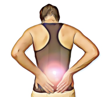
CUMBERLAND PHYSIOTHERAPY: Almost everyone will experience lower back and neck pain at some point in their lives, even if just in the form of a slight neck twinge after sleeping in an odd position. Spinal pain of the thoracic region is much less common, however, you might be surprised to know how important this part of the body is when it comes to pain and injury.

What is it? 

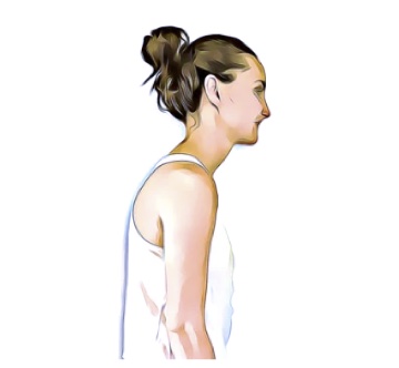
The thoracic refers to the part of the spine that is surrounded by the rib cage. It consists of 12 vertebrae with small, thick discs that sit between each of them. The thoracic spine isn’t an area that you might associate much with movement, however, this area can account for a surprising amount of flexibility, particularly in rotation.

With joint attachments both between each side of the 12 vertebrae and a rib on either side, the thoracic spine has almost more individual joints than you can count. If each of these

joints is not regularly moved through their full range they can tighten up and lose flexibility. This stiffness can become quite significant over time.

Why is it important?

Many people may not even notice this lack of movement, primarily because the neck and lower back provide much more range and can easily compensate for any loss of thoracic flexibility to complete everyday tasks.

When there is no movement occurring in the thoracic region, this means that the structures of the joints in other regions are pushed closer to their limits of range, particularly during rotation. This results in more compression and stress on these joints and the structures surrounding them, such as nerves, blood vessels and muscles.

Thoracic stiffness can be a significant risk factor for neck and lower back pain. This can also reduce the mobility of the chest wall, which can result in less efficient breathing mechanics and, in extreme cases, even reduced exercise tolerance.

How can physiotherapy help?

Your physiotherapist is able to assess your thoracic mobility and help you with treatments to improve your range, both with manual therapy and home exercises. They may even help improve your thoracic flexibility as part of a treatment plan for neck and lower back pain.

None of the information in this article is a replacement for proper medical advice. Always see a medical professional for advice on your injury. 

Five Physio Tips to Help Improve Your Health

CUMBERLAND PHYSIOTHERAPY: There’s no doubt that doing the right thing for your health can be difficult. Whether that involves eating healthier, exercising more, flossing your teeth or even doing your physio exercises, the best choice is not always the most comfortable. 

Once a habit has formed and become part of your routine, making the right decision for yourself can become a lot easier. Getting to that point is much easier said than done. Here are a few tips to help you reach your health goals.

1. Choose a friend to join you for accountability.

One way to guarantee that you show up for an early morning gym session is to have a friend who you partner with. You might be comfortable sleeping in for yourself, but not so much when you have someone else you’re letting down. Sharing difficult tasks with a friend can make them more enjoyable and you can also set up a competition between them if you need a little extra motivation.

2. Combine tasks with an activity that you do like. 

Love a true crime podcast? The gym can be the time to catch the latest episode or even stream your favourite show. Anything you do that helps to increase the enjoyment of the activity you’re avoiding is going to help you get started.

3. Batch tasks to be more efficient.

Are you trying to drink more water, floss and also complete your physio exercises? Why not do all three in the same sitting? Combining tasks can be a surprisingly effective use of time and a way to remind yourself to complete them.

3. Use positive reinforcement for good behaviour, rather than punishments. 

Many people will feel bad when they fail to reach their goals and try to restrict themselves in another area to compensate. It can be a much more effective motivation strategy to reward yourself after completing a task, rather than punish yourself for not doing it.

4. Get your foot in the door.

Putting your shoes on can be the hardest part of going for a run. If you’re struggling to complete a task, sometimes all you have to do is tell yourself you only need to do five minutes. Once you’ve started quite often you’ll want to do more, but setting small goals can be the trick to getting started.

Once a habit is in place, the easy part is to increase the intensity or length of time you’re doing it. It only takes 21 days of completing a task for a habit to be formed, where you will do it automatically as part of your routine. For more tips and tricks on how to reach your health goals, speak to your physiotherapist. 

What Is Chronic Ankle Instability


 Chronic ankle instability, as the name implies, is a chronic condition of instability affecting the ankle and it’s surrounding structures. It usually develops after a severe ankle sprain. However, some people are born with less stable ankles; these individuals are generally extra flexible throughout their bodies. Approximately 20% of ankle sprains lead to chronic ankle instability due to the resulting changes in ligament support, strength, postural control, muscle reaction time and sensation. 

What are the symptoms?

As well as being more susceptible to ankle sprains, people with chronic ankle instability may notice they are extra cautious during high-intensity activities, if running on uneven surfaces or when changing directions quickly. They may experience a sense of weakness or frequent ‘giving way’ when weight-bearing.

What are the causes?

The primary causes of this condition are ligament laxity, decreased muscle strength of the muscles surrounding the ankle and reduced proprioception. 

Following an ankle sprain, ligaments can be stretched and slightly weaker; in severe cases, they have torn altogether, leaving the ankle structurally weaker. Without full rehabilitation, the surrounding muscles also become weaker, and studies have shown that balance and sensation of the ankle can also be reduced. This means that the ankle is more likely to be injured again, creating a vicious cycle leading to further instability.

How can physiotherapy help?

Physiotherapy treatment for chronic ankle instability focuses on improving strength, control and balance with a variety of techniques. This approach can significantly improve ankle stability and reduce the risk of future sprains. Physiotherapists can help patients to regain confidence and get back to their best performance. 

In some cases, orthotic braces for support can be used. However, this can lead to dependence and further loss of strength and control if used unnecessarily. In cases of extreme ligament laxity or if physiotherapy fails, surgery to repair the damaged ligaments is considered. This is usually combined with a full physiotherapy rehabilitation program for greatest success.

If you don’t feel 100% confident with your ankle, come and have a chat with one of our physiotherapists to see if we can help improve your ankle stability. None of the information in this article is a replacement for proper medical advice. Always see a medical professional for advice on your individual injury. 

Five Reasons To See a Physiotherapist After Injury

 There is no doubt that the human body can be very resilient. Short of regenerating new limbs, our bodies are capable of recovering from large amounts of damage, including broken bones. With this in mind, many people are happy to let nature take it’s course following an injury, thinking that seeing a physiotherapist will only act to speed up already healing tissues.

The speed of recovery, however, is only one measure of healing and despite our bodies’ incredible capacity for repair; injury repair can be less than straightforward. Here are a few things about injury healing you may not have been aware of.

1. Scar Tissue is more likely to form without treatment. 

Scar tissue can cause ongoing pain and stiffness in skin, muscles and ligaments. Physiotherapy can prevent excessive scarring from forming through advice regarding movement, massage and other hands-on treatment. 

2. Your ability to sense the position of your body, known as proprioception, is often damaged after an injury and can be retrained. 

Impaired proprioception is a major factor in re-injury. If you’ve ever heard someone say “my knee/ankle/shoulder still doesn’t feel 100%” then this could be why. The good news is that with a specific exercise program, proprioception can be improved and recovered.

3. Once healing has finished, your body may not be exactly the same as before.

Following an injury, ligaments may be lax, joints may be stiffer and muscles are almost always weaker. While the pain may be gone, there might still be factors that need to be addressed to prevent more complicated issues in the future. 

4. You may have picked up some bad habits while waiting for the injury to heal. 

While in pain, we often change the way we do things, this can lead to the development of poor movement patterns and muscle imbalances. Even though the pain has gone, these new patterns can remain and create further problems down the road. 

5. Injuries don’t always heal completely.

 On rare occasions, injuries may not be able to heal completely on their own. The most serious example of this is a fracture that cannot heal if the bone is not kept still enough. Other factors that may prevent an injury from healing include poor circulation, diabetes, insufficient care of the injury and poor nutrition. 

Your physiotherapist can assess your injury and develop a treatment plan that will both restore you to the best possible function and prevent further injuries. None of the information in this article is a replacement for proper medical advice. Always see a medical professional for advice on your individual injury.  

Focus on Calf Tears

 What are they?

The calf muscles refer to a group of muscles at the back of the lower leg that act to point the foot away from the body and play an important role in walking and running. A tear or strain of these muscles occurs when some or all of the muscle fibres are torn or stretched. This is a common injury that can affect anyone from athletes to those with a more relaxed lifestyle.

How do they happen?

Calf tears are often caused by sudden, forceful movements or overuse of the calf muscles, leading to the rupture or strain of muscle fibres. Common mechanisms of injury are a quick take off during a sports match or simply going for a long walk when not accustomed. Factors that may increase the risk of a calf tear are previous calf tears that have not been fully rehabilitated, tight and weak calf muscles, poor balance and ill fitting footwear. 

What are the symptoms?

Typical symptoms of a calf tear are sharp pain over the site of the tear, especially with movement, swelling, bruising, and difficulty walking or standing. The severity of the injury can range from mild muscle strain to a complete tear, which will determine the appropriate treatment approach.

How can physiotherapy help?

The first step in managing calf tears is accurate diagnosis by a medical professional, who is able to rule out other conditions that might mimic a calf tear. They can determine the extent of the damage and create personalised treatment plans based on the patient’s specific needs. This ensures that the rehabilitation process addresses the root cause of the injury, leading to better outcomes.

Reducing pain and inflammation is important in the first one to two days following the injury. The muscle may need support during this time, depending on the severity. Over time as the swelling and inflammation subsides, your physiotherapist will help to address any factors that contributed to the injury such as muscle weakness or imbalance. Calf tears often lead to stiffness and limited range of motion in the affected leg.

Physiotherapists implement targeted stretching and range of motion exercises to restore flexibility and prevent the formation of scar tissue that may impede recovery. Gradually, the patient can regain the ability to move the calf muscle without pain or discomfort.

Rehabilitation past this point will progressively challenge the calf muscles without causing further damage. Strengthening these muscles not only aids in the healing process but also reduces the risk of future calf tears.

Proprioception, the body’s ability to sense its position in space, is crucial for balance and coordination. Physiotherapy includes specialised exercises that enhance proprioception and balance, reducing the likelihood of re-injury. This aspect of rehabilitation is especially important for athletes and active individuals who need to return to high-intensity activities safely.

None of the information in this article is a replacement for proper medical advice. Always see a medical professional for advice on your individual condition.

Surprising Skills That Improve With Age

 Many of us associate ageing with a decline of skills and quality of life. However, the truth is not as black and white as this. While certain skills and functions do tend to suffer as we age, surprisingly there are many abilities that actually improve as we grow older. 

Some studies have shown that happiness is U-shaped, proving that the mid-life crisis is real, with the ages between 40-60 holding the highest amount of stress and responsibility for adults. It seems that happiness increases steadily after this period, with the average 80 year old reporting themselves to be as happy as the average 20 year old. 

There is also evidence that your beliefs about ageing can act as a self-fulfilling prophecy. If you are positive about ageing, then your experience will be more positive than if you have negative beliefs about it. 

It’s not only good news about your mental health though. Studies have also shown that while your short-term memory might decline, other skills such as creativity can actually increase. One of the biggest reasons for a decline in skill and function is the disuse that comes with retirement from work. Keeping active and mentally stimulated can be enough to keep your skills up to speed. 

It has also been shown that confidence grows in both genders as we age. Making decisions becomes  easier as we know ourselves better and have a wealth of experience to draw on when a tricky situation arises. 

Certain physical skills such as strength and agility might decline, however it seems that other aspects of physical ability might increase, including endurance. There are many triathletes who are in their 70s, 80s, and unbelievably even in their 90s. In the absence of any serious disability, it might be the case that age is an excuse rather than an actual hindrance to being active. 

Verbal ability and vocabulary are also skills that improve as we age, which may explain why crosswords are so popular with the elderly. Making the most of your abilities at every age is important, so try not to let age be a barrier to trying new things and keeping active. 

Physiotherapists are dedicated to helping people stay active at any age and can help you with achieving your goals. Speak to your physiotherapist for more information.

Frozen Shoulder

 What is frozen shoulder?

Frozen Shoulder (also known as Adhesive Capsulitis) is a condition that affects the shoulder joint with no apparent (idiopathic) cause. It is characterised by severe pain and stiffness of the shoulder. The shoulder capsule thickens due to lack of synovial fluid, which leads to tightness and stiffness. Frozen shoulder occurs most frequently between the ages of 40-60 years old and affects more women than men. 

How does it happen?

The cause of Frozen Shoulder is still unknown, however some risk factors have been identified that increase your chances of developing the condition including diabetes mellitus, stroke, shoulder injuries and immobilisation.

The hallmark sign of Frozen Shoulder is being unable to move your shoulder even with the help of other people. This is particularly noticeable when moving the arm away from the body. Physicians may ask you to undergo investigations such as X-ray and MRI, however frozen shoulder doesn’t tend to show up on imaging. 

What are the symptoms?

Frozen Shoulder has three stages:

1. Freezing (Painful stage) – lasts from six weeks to nine months; patient has a slow onset of pain (usually pain at rest), and the shoulder starts to experience limitation of motion

2. Frozen (Adhesive stage) – lasts from four to six months; pain begins to diminish (the shoulder is still usually painful with movement), of the shoulder getting stiffer, and activities of daily living are affected).

3. Thawing (Recovery stage) – last six months to two years; shoulder’s normal range of motion is slowly returning to normal

How can physiotherapy help?

Though Frozen Shoulder is a self-limiting condition, an important part of physiotherapy management is the prevention of related neck and shoulder issues secondary to the original problem. As you can imagine, frozen shoulder can be extremely debilitating and many adaptations occur in the surrounding musculature. Physiotherapy also acts to reduce pain throughout phases one and two, while restoring joint movement as the joint moves through the painful phase. This is done through a variety of treatments, including heat, stretching, joint mobilisation, range of motion exercises to improve shoulder’s motion and resistance exercise to strengthen, you will also be given a home exercise program. Recovery time varies with every patient. If you’re working hard to regain your normal shoulder function, you’ll be rewarded with a faster recovery.

Medical management includes medications and corticosteroids, joint manipulation while under anaesthetic and/or surgery, however at this point nothing has come close to providing a consistent and simple cure.

None of the information in this article is a replacement for proper medical advice. Always see a medical professional for advice on your individual condition. 

Tips for Exercising when You have Pain

 One of the most challenging aspects of living with an injury or chronic pain is how it can quickly impact your exercise routine. If you have been working towards a fitness or weight goal, this can be extremely demoralising. Here are a few tips that can help to keep you on track while you recover. Staying as active as possible during this time can mean you’re in the best position to reach your goals again once your injury has healed. 

1. Try a new activity. 

When injury strikes, it can be tempting to stop exercising altogether and rest while you recover. An injury can be frustrating, but it can also be an opportunity to try out a different sport. If you’re a runner with an ankle injury, you can keep up your fitness by swimming instead. Cycling can be an excellent option for people for dealing with knee pain, and if you’re a swimmer with shoulder pain, maybe switch to running for a while. Check with your physiotherapist for some ideas to keep you moving. 

2. Exercise within your limits. 

If you’re getting pain at 5km, this doesn’t always mean you should give up running altogether. Your physiotherapist can help you monitor your symptoms carefully and plan an exercise routine that keeps your fitness up while reducing symptom flare-ups. Staying as active as possible throughout your recovery can also mean that you a better placed to get back to your best performance once symptoms reside

3. Take the opportunity to improve your footwear and equipment. 

Injury and pain can be a great prompt to look at your equipment and technique . For example, with hip and knee pain, the type of shoes you wear can have a significant difference. Often pain has more than once cause, with technique and equipment more often than not having a substantial impact on the stress placed on your body. Your physiotherapist is an excellent source of advice in this area, don’t hesitate to ask for an assessment. 

4. Take to the water

Hydrotherapy has long been used to help patients with joint pain or muscle weakness exercise. The water helps reduce joint stress and provide extra sensory input that can reduce pain. Exercising in water can be especially helpful for sufferers of chronic pain or those who have pain with weight-bearing. Speak to your physio for a hydrotherapy program if you’re not sure how to approach exercise in water. 

Our physiotherapists are happy to discuss your condition with you and share their tips to help you stay pain-free. None of the information in this article is a replacement for proper medical advice. Always see a medical professional for advice on your condition.

Common myths around MRI and Back pain

CUMBERLAND PHYSIOTHERAPY PARRAMATTA:If you’ve been unlucky enough to suffer from chronic or ongoing back pain, chances are that at some stage, you’ve had other investigations to capture what is happening inside your spine. Magnetic resonance imaging (MRI) is an incredible technology that can provide essential insights into tissues that otherwise can’t be seen.

Unfortunately, there are some common misconceptions around what that information means, which can sometimes be unhelpful and even harmful to recovery. Here are a few things you might not realize that can help you understand your MRI:

Not all tissues show up on every scan.

Muscle, fascia, or other irritable tissues may not show up on your scan, meaning you may experience a lot of pain but have a completely normal scan. Not all parts of your anatomy will show up from every scan’s perspective and some tissue changes might only be evident in certain postures.

Age-related changes are normal and may not be related to your pain.

Results such as arthritis, disc bulges, and small tears can be concerning. However, it’s helpful to note that people who have no painful symptoms can have similar, if not more, age-related changes, and these signs may not be related to your pain at all.

Severe pain may not be related to severe changes on MRI.

Pain is a complex experience, influenced by multiple factors including the sensitivity of the tissues involved, what the brain believes about the pain and what it means, and how long it has been present. MRI is helpful for ruling out severe pathology such as infection, fracture, or malignancy; however, it is not always an accurate guide to the source of symptoms. The results of an MRI should always be taken into account as a small part of a bigger clinical picture when guiding treatment.

Talk to your physiotherapist for advice on any imaging results you have and what they might mean for your treatment and prognosis. None of the information in this article is a replacement for professional medical advice. Always consult a medical professional for advice on your condition.

Fibula Fractures

CUMBERLAND PHYSIOTHERAPY PARRAMATTA:

What are they?

The lower part of the leg, from the knee down to the ankle, consists of two long bones that sit side by side: the thicker Tibia and the thinner Fibula. The bones are joined together by thick fibrous connective tissue called a “syndesmosis” and are firmly adhered to each other with just a small amount of movement between them, allowing for a small amount of rotation of the ankle.

A fracture of the fibula occurs when the bony tissue is disrupted or broken. It is a common injury and can occur at any part of the bone, depending on the mechanism of injury or the state of the bone.

How does it happen?

A fracture of any bone can occur when the force applied to any point exceeds the strength of the tissue at that point. However, as with all fractures, there are common patterns that are seen based on structural points of weakness in the bone and common patterns of movement. A few common ways that the fibula is broken are;

Blunt force: 

If something hits the bone hard enough, it will break on impact. This could include being hit by a ball hard enough or being hit by a car, as this is the site where a car’s bumper would reach. Impacts like this that have enough force will often break both the Tibia and the Fibula at the same time. Skiing accidents where skis hit something suddenly or get stuck can also cause the bones to break at the level of the ski boots.

Ankle Sprain: 

When it comes to the fibula, the most common reason for the bone to be broken is during a severe ankle sprain. The ligaments that attach the outside of the foot to the fibula are so strong that when you twist your ankle badly enough, sometimes it is the bone that breaks. This is one of the most commonly missed injuries, partly because the fibula is not a weight-bearing bone. This means that after the initial pain and swelling have subsided, you can still walk on your foot without pain stopping you. It is important to have any severe ankle sprains imaged by X-ray to rule out any fibula fractures.

What are the symptoms?

In some cases, the symptoms of a fibula fracture will be unmistakable, with severe pain. Sometimes the skin will be broken and there will be bleeding. If the bone has been moved from its usual position, there will be a deformity under the skin. For smaller, displaced fractures, there will be pain over the bony aspects and a constant, deep pain that is worse when weight-bearing.

What is the treatment?

Physiotherapists are often the first to notice fractures caused by ankle sprains. Once a fracture has been confirmed, your medical team will decide on the best course of action to allow the bones to heal. This might include surgery to pin the bones together, casting or the use of a moon boot. Following a period of immobilisation, your physiotherapist can help you rehabilitate the surrounding tissues. This will include muscle strengthening, joint mobilisation, balance and control retraining, and a stretching program.

None of the information in this article is a replacement for proper medical advice. Always seek medical professional advice for your condition.