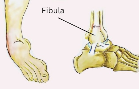
Focus on Calf Tears

 What are they?

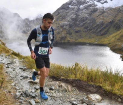
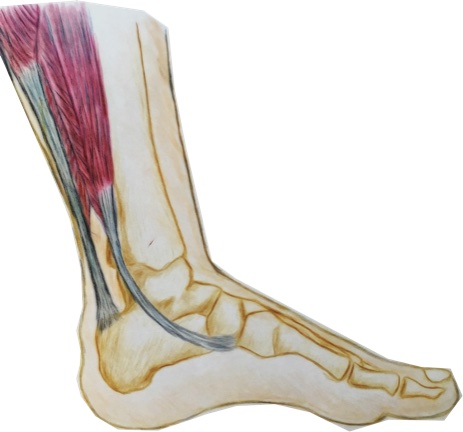
The calf muscles refer to a group of muscles at the back of the lower leg that act to point the foot away from the body and play an important role in walking and running. A tear or strain of these muscles occurs when some or all of the muscle fibres are torn or stretched. This is a common injury that can affect anyone from athletes to those with a more relaxed lifestyle.

How do they happen?

Calf tears are often caused by sudden, forceful movements or overuse of the calf muscles, leading to the rupture or strain of muscle fibres. Common mechanisms of injury are a quick take off during a sports match or simply going for a long walk when not accustomed. Factors that may increase the risk of a calf tear are previous calf tears that have not been fully rehabilitated, tight and weak calf muscles, poor balance and ill fitting footwear. 

What are the symptoms?

Typical symptoms of a calf tear are sharp pain over the site of the tear, especially with movement, swelling, bruising, and difficulty walking or standing. The severity of the injury can range from mild muscle strain to a complete tear, which will determine the appropriate treatment approach.

How can physiotherapy help?

The first step in managing calf tears is accurate diagnosis by a medical professional, who is able to rule out other conditions that might mimic a calf tear. They can determine the extent of the damage and create personalised treatment plans based on the patient’s specific needs. This ensures that the rehabilitation process addresses the root cause of the injury, leading to better outcomes.

Reducing pain and inflammation is important in the first one to two days following the injury. The muscle may need support during this time, depending on the severity. Over time as the swelling and inflammation subsides, your physiotherapist will help to address any factors that contributed to the injury such as muscle weakness or imbalance. Calf tears often lead to stiffness and limited range of motion in the affected leg.

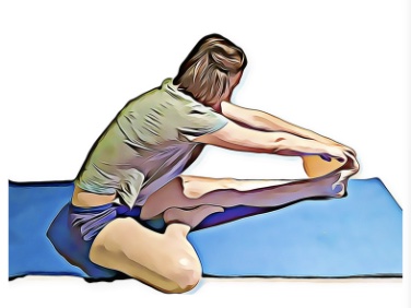
Physiotherapists implement targeted stretching and range of motion exercises to restore flexibility and prevent the formation of scar tissue that may impede recovery. Gradually, the patient can regain the ability to move the calf muscle without pain or discomfort.

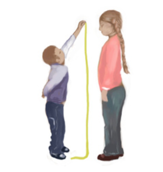
Rehabilitation past this point will progressively challenge the calf muscles without causing further damage. Strengthening these muscles not only aids in the healing process but also reduces the risk of future calf tears.

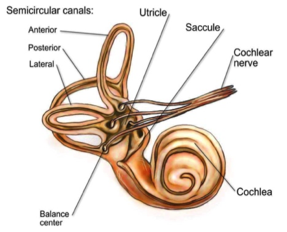
Proprioception, the body’s ability to sense its position in space, is crucial for balance and coordination. Physiotherapy includes specialised exercises that enhance proprioception and balance, reducing the likelihood of re-injury. This aspect of rehabilitation is especially important for athletes and active individuals who need to return to high-intensity activities safely.

None of the information in this article is a replacement for proper medical advice. Always see a medical professional for advice on your individual condition.

Surprising Skills That Improve With Age

 Many of us associate ageing with a decline of skills and quality of life. However, the truth is not as black and white as this. While certain skills and functions do tend to suffer as we age, surprisingly there are many abilities that actually improve as we grow older. 

Some studies have shown that happiness is U-shaped, proving that the mid-life crisis is real, with the ages between 40-60 holding the highest amount of stress and responsibility for adults. It seems that happiness increases steadily after this period, with the average 80 year old reporting themselves to be as happy as the average 20 year old. 

There is also evidence that your beliefs about ageing can act as a self-fulfilling prophecy. If you are positive about ageing, then your experience will be more positive than if you have negative beliefs about it. 

It’s not only good news about your mental health though. Studies have also shown that while your short-term memory might decline, other skills such as creativity can actually increase. One of the biggest reasons for a decline in skill and function is the disuse that comes with retirement from work. Keeping active and mentally stimulated can be enough to keep your skills up to speed. 

It has also been shown that confidence grows in both genders as we age. Making decisions becomes  easier as we know ourselves better and have a wealth of experience to draw on when a tricky situation arises. 

Certain physical skills such as strength and agility might decline, however it seems that other aspects of physical ability might increase, including endurance. There are many triathletes who are in their 70s, 80s, and unbelievably even in their 90s. In the absence of any serious disability, it might be the case that age is an excuse rather than an actual hindrance to being active. 

Verbal ability and vocabulary are also skills that improve as we age, which may explain why crosswords are so popular with the elderly. Making the most of your abilities at every age is important, so try not to let age be a barrier to trying new things and keeping active. 

Physiotherapists are dedicated to helping people stay active at any age and can help you with achieving your goals. Speak to your physiotherapist for more information.

Frozen Shoulder

 What is frozen shoulder?

Frozen Shoulder (also known as Adhesive Capsulitis) is a condition that affects the shoulder joint with no apparent (idiopathic) cause. It is characterised by severe pain and stiffness of the shoulder. The shoulder capsule thickens due to lack of synovial fluid, which leads to tightness and stiffness. Frozen shoulder occurs most frequently between the ages of 40-60 years old and affects more women than men. 

How does it happen?

The cause of Frozen Shoulder is still unknown, however some risk factors have been identified that increase your chances of developing the condition including diabetes mellitus, stroke, shoulder injuries and immobilisation.

The hallmark sign of Frozen Shoulder is being unable to move your shoulder even with the help of other people. This is particularly noticeable when moving the arm away from the body. Physicians may ask you to undergo investigations such as X-ray and MRI, however frozen shoulder doesn’t tend to show up on imaging. 

What are the symptoms?

Frozen Shoulder has three stages:

1. Freezing (Painful stage) – lasts from six weeks to nine months; patient has a slow onset of pain (usually pain at rest), and the shoulder starts to experience limitation of motion

2. Frozen (Adhesive stage) – lasts from four to six months; pain begins to diminish (the shoulder is still usually painful with movement), of the shoulder getting stiffer, and activities of daily living are affected).

3. Thawing (Recovery stage) – last six months to two years; shoulder’s normal range of motion is slowly returning to normal

How can physiotherapy help?

Though Frozen Shoulder is a self-limiting condition, an important part of physiotherapy management is the prevention of related neck and shoulder issues secondary to the original problem. As you can imagine, frozen shoulder can be extremely debilitating and many adaptations occur in the surrounding musculature. Physiotherapy also acts to reduce pain throughout phases one and two, while restoring joint movement as the joint moves through the painful phase. This is done through a variety of treatments, including heat, stretching, joint mobilisation, range of motion exercises to improve shoulder’s motion and resistance exercise to strengthen, you will also be given a home exercise program. Recovery time varies with every patient. If you’re working hard to regain your normal shoulder function, you’ll be rewarded with a faster recovery.

Medical management includes medications and corticosteroids, joint manipulation while under anaesthetic and/or surgery, however at this point nothing has come close to providing a consistent and simple cure.

None of the information in this article is a replacement for proper medical advice. Always see a medical professional for advice on your individual condition. 

Tips for Exercising when You have Pain

 One of the most challenging aspects of living with an injury or chronic pain is how it can quickly impact your exercise routine. If you have been working towards a fitness or weight goal, this can be extremely demoralising. Here are a few tips that can help to keep you on track while you recover. Staying as active as possible during this time can mean you’re in the best position to reach your goals again once your injury has healed. 

1. Try a new activity. 

When injury strikes, it can be tempting to stop exercising altogether and rest while you recover. An injury can be frustrating, but it can also be an opportunity to try out a different sport. If you’re a runner with an ankle injury, you can keep up your fitness by swimming instead. Cycling can be an excellent option for people for dealing with knee pain, and if you’re a swimmer with shoulder pain, maybe switch to running for a while. Check with your physiotherapist for some ideas to keep you moving. 

2. Exercise within your limits. 

If you’re getting pain at 5km, this doesn’t always mean you should give up running altogether. Your physiotherapist can help you monitor your symptoms carefully and plan an exercise routine that keeps your fitness up while reducing symptom flare-ups. Staying as active as possible throughout your recovery can also mean that you a better placed to get back to your best performance once symptoms reside

3. Take the opportunity to improve your footwear and equipment. 

Injury and pain can be a great prompt to look at your equipment and technique . For example, with hip and knee pain, the type of shoes you wear can have a significant difference. Often pain has more than once cause, with technique and equipment more often than not having a substantial impact on the stress placed on your body. Your physiotherapist is an excellent source of advice in this area, don’t hesitate to ask for an assessment. 

4. Take to the water

Hydrotherapy has long been used to help patients with joint pain or muscle weakness exercise. The water helps reduce joint stress and provide extra sensory input that can reduce pain. Exercising in water can be especially helpful for sufferers of chronic pain or those who have pain with weight-bearing. Speak to your physio for a hydrotherapy program if you’re not sure how to approach exercise in water. 

Our physiotherapists are happy to discuss your condition with you and share their tips to help you stay pain-free. None of the information in this article is a replacement for proper medical advice. Always see a medical professional for advice on your condition.

Common myths around MRI and Back pain

CUMBERLAND PHYSIOTHERAPY PARRAMATTA:If you’ve been unlucky enough to suffer from chronic or ongoing back pain, chances are that at some stage, you’ve had other investigations to capture what is happening inside your spine. Magnetic resonance imaging (MRI) is an incredible technology that can provide essential insights into tissues that otherwise can’t be seen.

Unfortunately, there are some common misconceptions around what that information means, which can sometimes be unhelpful and even harmful to recovery. Here are a few things you might not realize that can help you understand your MRI:

Not all tissues show up on every scan.

Muscle, fascia, or other irritable tissues may not show up on your scan, meaning you may experience a lot of pain but have a completely normal scan. Not all parts of your anatomy will show up from every scan’s perspective and some tissue changes might only be evident in certain postures.

Age-related changes are normal and may not be related to your pain.

Results such as arthritis, disc bulges, and small tears can be concerning. However, it’s helpful to note that people who have no painful symptoms can have similar, if not more, age-related changes, and these signs may not be related to your pain at all.

Severe pain may not be related to severe changes on MRI.

Pain is a complex experience, influenced by multiple factors including the sensitivity of the tissues involved, what the brain believes about the pain and what it means, and how long it has been present. MRI is helpful for ruling out severe pathology such as infection, fracture, or malignancy; however, it is not always an accurate guide to the source of symptoms. The results of an MRI should always be taken into account as a small part of a bigger clinical picture when guiding treatment.

Talk to your physiotherapist for advice on any imaging results you have and what they might mean for your treatment and prognosis. None of the information in this article is a replacement for professional medical advice. Always consult a medical professional for advice on your condition.

Fibula Fractures

CUMBERLAND PHYSIOTHERAPY PARRAMATTA:

What are they?

The lower part of the leg, from the knee down to the ankle, consists of two long bones that sit side by side: the thicker Tibia and the thinner Fibula. The bones are joined together by thick fibrous connective tissue called a “syndesmosis” and are firmly adhered to each other with just a small amount of movement between them, allowing for a small amount of rotation of the ankle.

A fracture of the fibula occurs when the bony tissue is disrupted or broken. It is a common injury and can occur at any part of the bone, depending on the mechanism of injury or the state of the bone.

How does it happen?

A fracture of any bone can occur when the force applied to any point exceeds the strength of the tissue at that point. However, as with all fractures, there are common patterns that are seen based on structural points of weakness in the bone and common patterns of movement. A few common ways that the fibula is broken are;

Blunt force: 

If something hits the bone hard enough, it will break on impact. This could include being hit by a ball hard enough or being hit by a car, as this is the site where a car’s bumper would reach. Impacts like this that have enough force will often break both the Tibia and the Fibula at the same time. Skiing accidents where skis hit something suddenly or get stuck can also cause the bones to break at the level of the ski boots.

Ankle Sprain: 

When it comes to the fibula, the most common reason for the bone to be broken is during a severe ankle sprain. The ligaments that attach the outside of the foot to the fibula are so strong that when you twist your ankle badly enough, sometimes it is the bone that breaks. This is one of the most commonly missed injuries, partly because the fibula is not a weight-bearing bone. This means that after the initial pain and swelling have subsided, you can still walk on your foot without pain stopping you. It is important to have any severe ankle sprains imaged by X-ray to rule out any fibula fractures.

What are the symptoms?

In some cases, the symptoms of a fibula fracture will be unmistakable, with severe pain. Sometimes the skin will be broken and there will be bleeding. If the bone has been moved from its usual position, there will be a deformity under the skin. For smaller, displaced fractures, there will be pain over the bony aspects and a constant, deep pain that is worse when weight-bearing.

What is the treatment?

Physiotherapists are often the first to notice fractures caused by ankle sprains. Once a fracture has been confirmed, your medical team will decide on the best course of action to allow the bones to heal. This might include surgery to pin the bones together, casting or the use of a moon boot. Following a period of immobilisation, your physiotherapist can help you rehabilitate the surrounding tissues. This will include muscle strengthening, joint mobilisation, balance and control retraining, and a stretching program.

None of the information in this article is a replacement for proper medical advice. Always seek medical professional advice for your condition.

Tips for Optimal Stretching Results

 How do you stretch properly?

CUMBERLAND PHYSIOTHERAPY PARRAMATTA: While there are many benefits to be gained from effective stretching, employing an improper technique can actually result in injury. Here are some tips and guidelines for you to follow when stretching: 

·Warm up first with an activity such as walking (at least 5 -10 minutes).

·Stretches should be pain-free. You should only feel tension or a tight feeling when stretching. If you feel a sharp pain, this means you’re stretching too far and should relax a little. 

·Don’t hold your breath while stretching; keep breathing to ensure the muscle tissues remain oxygenated during the stretch.

·Stretch both sides. But, if one muscle is tighter than the other, focus on it more until they’re both in the same range.

·Avoid bouncing at the end of the stretch because it may lead to injury.

·Hold stretches for 20-30 seconds, if not longer. A 10 second stretch is not always enough to achieve a lasting effect.

·Repeat the stretch 3-5 times with intermittent rest periods in between.

·Make sure your body is aligned properly and observe good posture.

When to Stretch?

Traditionally, stretching has been encouraged before and after any kind of physical activity. However, recent studies show that there is no discerning difference between whether you stretch before exercise/sports or not, both-in terms of performance and injury prevention. In some cases, researchers say that it’s okay to omit the pre-event stretch since the post-event stretch is much more beneficial. 

Some of our everyday postures and activities can lead to certain muscles becoming tight while the others sit in a lengthened position. These habitual postures that lead to muscle imbalances then become more and more difficult to correct over time. Many therapists will suggest that stretching all muscles generically isn’t as beneficial as identifying which of your muscles are abnormally tight and developing a targeted stretching regime. 

Talk to your physiotherapist for advice regarding the best stretching program for your body type, posture and activity levels. 

Positional Vertigo

CUMBERLAND PHYSIOTHERAPY PARRAMATTA:

What is it?

Vertigo, the feeling that you are moving even though you aren’t, is an unpleasant yet common experience caused by a variety of conditions. It may surprise you to learn that in some cases your physiotherapist is actually able to treat vertigo.

The process of telling whether we are moving or still involves many parts of the nervous system, including the inner ear. The vestibulocochlear nerve sends information about head movement to the brain, where it is processed. However, certain conditions can disrupt this process, causing the brain to perceive movement when there is none. Physiotherapists may be able to help with a specific type of vertigo called Benign Paroxysmal Positional Vertigo (BPPV).

What are the symptoms?

BPPV, also known as positional vertigo, causes dizziness only when the head moves in certain positions or directions. People with BPPV often experience dizziness and nausea when rolling over in bed or looking upwards, as well as lightheadedness and disturbance of balance. While BPPV can occur for no reason, it is often seen after a recent head trauma, respiratory infection, or airplane travel, as these conditions can disrupt the inner ear’s normal function.

How does it happen?

The symptoms of BPPV can be explained by a disruption in the signal sent by the semi-circular canals of the inner ear to the brain. These canals are positioned in different directions and filled with fluid. As the head moves, the fluid in each canal moves differently, depending on the head’s orientation. Receptors pick up this movement direction and speed, sending the message to the brain. However, sometimes small calcium crystals in the utricle, where the three semi-circular canals meet, can become dislodged and move into the semi-circular canals, disrupting the fluid and obscuring the messages to the brain.

What is the treatment?

If your doctor has diagnosed you with BPPV, they or your physiotherapist can show you a series of movements to help dislodge the calcium crystals and move them away from the semi-circular canal. You may also be asked to perform exercises to prevent the crystals from returning. Usually, only one or two treatments are needed for symptom resolution, although some cases may require more.

If you think you have vertigo, it is important to be assessed by a medical professional, as there are many conditions that can cause these symptoms and correct diagnosis is necessary before treatment. None of the information in this article is a replacement for proper medical advice. Always see a medical professional for advice on your condition.

Growing Pains

CUMBERLAND PHYSIOTHERAPY PARRAMATTA:

Are growing pains real?

The short answer is that yes, growing pains are a real and usually harmless part of childhood. Though poorly understood, they are recognized as a common phenomenon occurring most often between the ages of 3 and 12. The pain is commonly felt in both legs, particularly at night with no clear cause of pain. As yet no one is able to explain why they happen but growing pains are thought to be a normal response of a growing body as it adapts to new heights, sizes, strengths and skills. 

Does this mean I can ignore my child’s pain?

Not so fast. While growing pains are harmless and usually transient, there are many childhood illnesses and conditions that do require professional assessment and, if left untreated, can cause serious harm. These include but are not limited to; Juvenile arthritis, childhood cancers (which often first present as knee or jaw pain), developmental hip dysplasia (abnormality of the hip joint), Perthes disease and a variety of other musculoskeletal disorders. 

While it’s true that children are generally more resilient and heal well, they are also 

vulnerable to injuries just like adults. All serious strains and sprains should be rehabilitated correctly to ensure no long-term problems occur down the track. Many childhood pains can also be relieved with physiotherapy in the short term even if the child will eventually grow out of the pain. 

How can I tell if pain is abnormal?

Unfortunately, unless you are a trained professional you won’t be able to tell. If there is any doubt in your mind always contact a physiotherapist or doctor. Many clinicians have great respect for a parent’s intuition and acknowledge that parents are usually very good at knowing if something is wrong with their child. 

Even if you’re sure nothing is wrong, there are a few signs and symptoms that you should take particular notice of. pain that is severe, pain that occurs suddenly without an obvious cause, pain that is one sided, pain that affects your child’s activity levels, causes a limp or is associated with signs of general illness/fever. 

Constant, severe and unrelenting pain is a serious sign that should be investigated at any age. If you’re worried, the first step is to consult a physiotherapist or general practitioner. 

None of the information in this article is a replacement for proper medical advice. Always see a medical professional for advice on your injury. 


Physio Tips for Comfortable Travel

CUMBERLAND PHYSIOTHERAPY PARRAMATTA: Travel can be one of the most enjoyable aspects of life, however if a trip away comes with the risk of increased pain and soreness, the excitement can be thoroughly diminished. Many people will even avoid travel altogether if their symptoms are exacerbated too much by long seated periods. If you are unlucky enough to fall into this category, here are a few tips from your physiotherapist that might help make long trips a little more comfortable.

Lower back pain is the chief complaint of most travellers. To avoid low back pain, try the following tips:

1) Adjust the seat angle. 

If you are able, tilt the seat slightly down towards your feet, not towards your hips. Ideally, your knees should be level with your hips, or even slightly lower. A footrest to lift your knees can also help.

2) Lift the height of the whole seat. This will assist with tip #1 by lifting the whole body away from the floor. When your hips are less bent, it is easier for your back to sit comfortably in an upright position.

3) Use a lower back support. 

Many cars nowadays come equipped with in-built lumbar supports. Often this support is a generic design and not fitted for your specific shape or height. A proper lumbar pillow, or even a rolled towel in the small of the back, can be more effective in maintaining the natural arch of your spine. This relieves pressure through your intervertebral discs, spinal ligaments, and spinal muscles.

Neck discomfort is easier to improve when tips for lower back are implemented first. Some very easy tips to reduce neck pain are as follows:

1) Use a travel pillow. 

If travelling on an airplane, try wearing the pillow backwards or sideways. This assists in supporting the head in a slightly better position when you rest or sleep, which helps to reduce the stress placed on the muscles, ligaments and joints within the neck.

2) Every hour, perform 10 chin tucks. 

This is easiest to perform correctly if you sit tall and press your chin straight backwards, lengthening the back of your neck. Do not tuck the chin to the chest. This exercise stretches the small postural muscles at the base of the skull, relaxing them.

The tips above are only suggestions. If you find they are helpful, continue performing them throughout your travels. However, if any of the above tips cause you pain or discomfort, it is advisable to seek your physiotherapist’s opinion.